Copyright
©The Author(s) 2015.
World J Hepatol. Jul 18, 2015; 7(14): 1884-1893
Published online Jul 18, 2015. doi: 10.4254/wjh.v7.i14.1884
Published online Jul 18, 2015. doi: 10.4254/wjh.v7.i14.1884
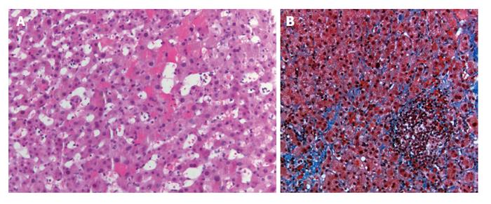
Figure 1 Hemotoxylin-eosin stains of a core liver biopsy from a patient with venous outflow obstruction shows hemorrhage within sinusoidal spaces (A) as well as evidence of sinusoidal fibrosis on a trichrome stain (B).
- Citation: Sarwar A, Ahn E, Brennan I, Brook OR, Faintuch S, Malik R, Khwaja K, Ahmed M. Utility of liver biopsy in predicting clinical outcomes after percutaneous angioplasty for hepatic venous obstruction in liver transplant patients. World J Hepatol 2015; 7(14): 1884-1893
- URL: https://www.wjgnet.com/1948-5182/full/v7/i14/1884.htm
- DOI: https://dx.doi.org/10.4254/wjh.v7.i14.1884


