Copyright
©The Author(s) 2015.
World J Hepatol. Jun 18, 2015; 7(11): 1460-1483
Published online Jun 18, 2015. doi: 10.4254/wjh.v7.i11.1460
Published online Jun 18, 2015. doi: 10.4254/wjh.v7.i11.1460
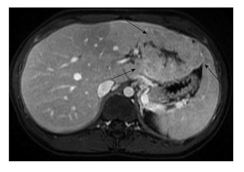
Figure 7 Magnetic resonance imaging of hepatocellular carcinoma in 19-year-old female.
Post contrast liver magnetic resonance imaging in portal venous phase shows a large mass (arrows) arising from the left lobe of a liver without cirrhosis. This lesion that has some imaging similarities with focal nodular hyperplasia, corresponded to fibrolamellar carcinoma on pathologic analysis.
- Citation: Yeh MM, Yeung RS, Apisarnthanarax S, Bhattacharya R, Cuevas C, Harris WP, Hon TLK, Padia SA, Park JO, Riggle KM, Daoud SS. Multidisciplinary perspective of hepatocellular carcinoma: A Pacific Northwest experience. World J Hepatol 2015; 7(11): 1460-1483
- URL: https://www.wjgnet.com/1948-5182/full/v7/i11/1460.htm
- DOI: https://dx.doi.org/10.4254/wjh.v7.i11.1460


