Copyright
©The Author(s) 2015.
World J Hepatol. Jun 18, 2015; 7(11): 1460-1483
Published online Jun 18, 2015. doi: 10.4254/wjh.v7.i11.1460
Published online Jun 18, 2015. doi: 10.4254/wjh.v7.i11.1460
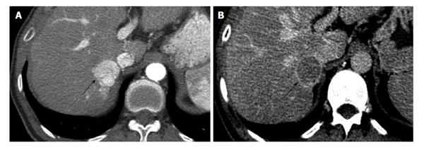
Figure 5 Computed tomography of hepatocellular carcinoma in 48-year-old male with hepatitis C.
A: Arterial phase contrast enhanced CT of the liver shows a strongly enhancing mass (arrow) in the right lobe, adjacent to the IVC. B: The same lesion (arrow) washes-out of contrast on the delayed phase and shows a thin capsule, this is diagnostic for HCC and corresponds to LI-RADS category 5. HCC: Hepatocellular carcinoma; CT: Computed tomography; LI-RADS: Liver imaging reporting and data system; IVC: Inferior vena cava.
- Citation: Yeh MM, Yeung RS, Apisarnthanarax S, Bhattacharya R, Cuevas C, Harris WP, Hon TLK, Padia SA, Park JO, Riggle KM, Daoud SS. Multidisciplinary perspective of hepatocellular carcinoma: A Pacific Northwest experience. World J Hepatol 2015; 7(11): 1460-1483
- URL: https://www.wjgnet.com/1948-5182/full/v7/i11/1460.htm
- DOI: https://dx.doi.org/10.4254/wjh.v7.i11.1460


