Copyright
©The Author(s) 2015.
World J Hepatol. Jun 18, 2015; 7(11): 1460-1483
Published online Jun 18, 2015. doi: 10.4254/wjh.v7.i11.1460
Published online Jun 18, 2015. doi: 10.4254/wjh.v7.i11.1460
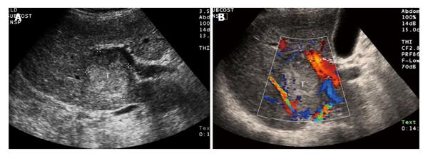
Figure 3 A 68-year-old male with cirrhosis and surgically proven hepatocellular carcinoma.
A: Thirty-four seconds after intravenous injection of ultrasound contrast (microbubbles) there is tumor (T with dashed line) enhancement; B: One and half minutes after injection the tumor (T with dashed line) is washing out of contrast. The images on the right side are a conventional sonogram (non-contrasted) of the lesion. The image on the left is a pulse inversion harmonics ultrasound for better visualization of ultrasound contrast media.
- Citation: Yeh MM, Yeung RS, Apisarnthanarax S, Bhattacharya R, Cuevas C, Harris WP, Hon TLK, Padia SA, Park JO, Riggle KM, Daoud SS. Multidisciplinary perspective of hepatocellular carcinoma: A Pacific Northwest experience. World J Hepatol 2015; 7(11): 1460-1483
- URL: https://www.wjgnet.com/1948-5182/full/v7/i11/1460.htm
- DOI: https://dx.doi.org/10.4254/wjh.v7.i11.1460


