Copyright
©The Author(s) 2015.
World J Hepatol. Jun 18, 2015; 7(11): 1460-1483
Published online Jun 18, 2015. doi: 10.4254/wjh.v7.i11.1460
Published online Jun 18, 2015. doi: 10.4254/wjh.v7.i11.1460
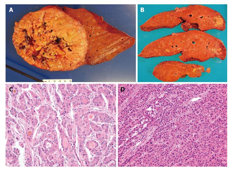
Figure 1 Pathology of classical hepatocellular carcinoma.
A: Gross photo of a well circumscribed, soft, yellowish to tan, and lobulated hepatocellular carcinoma (HCC) in a background of non-cirrhotic liver; B: Gross photo of a yellow and greenish, soft and lobulated HCC in a background of cirrhotic liver; C: Microphotos of HCC showing the pseudoacinar and pseudoglandular patterns, some containg the yellowish bile within the pseudoglandular structure with increased nuclear sizes; D: Microphotos of HCC showing thickened trabeculi, with increased unpaired arteries. Notice there are no normal structures present, i.e., portal tracts.
- Citation: Yeh MM, Yeung RS, Apisarnthanarax S, Bhattacharya R, Cuevas C, Harris WP, Hon TLK, Padia SA, Park JO, Riggle KM, Daoud SS. Multidisciplinary perspective of hepatocellular carcinoma: A Pacific Northwest experience. World J Hepatol 2015; 7(11): 1460-1483
- URL: https://www.wjgnet.com/1948-5182/full/v7/i11/1460.htm
- DOI: https://dx.doi.org/10.4254/wjh.v7.i11.1460


