Copyright
©2014 Baishideng Publishing Group Inc.
World J Hepatol. Aug 27, 2014; 6(8): 580-595
Published online Aug 27, 2014. doi: 10.4254/wjh.v6.i8.580
Published online Aug 27, 2014. doi: 10.4254/wjh.v6.i8.580
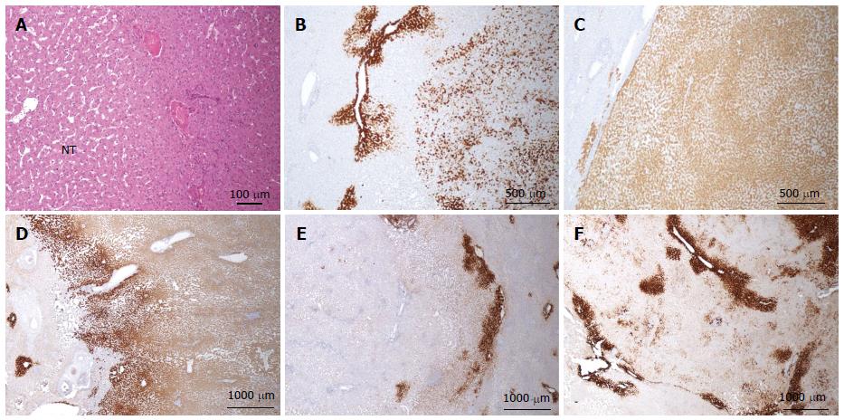
Figure 15 β-catenin activated, inflammatory hepatocellular adenoma.
A-C: Woman born in 1971; liver hemorrhage. No oral contraceptives. BMI 20.4 kg/m2. Imaging 5 nodules, largest 8 cm. Segmentectomy III, VI, VIII 2007. A: HE - large arteries at the periphery of the nodule. Mild sinusoidal dilatation in the non tumoral liver (NT). B: Abnormal patchy GS staining in one nodule. C: Abnormal homogeneous glutamine synthase (GS) staining in another nodule. D-F: Woman born in 1959; oral contraceptives for 21 years. BMI 21.8 kg/m2. Abnormal liver function tests. Imaging: 3 nodules, largest 10 cm. Right hepatectomy 2005. In the three nodules, GS staining is different but definitely abnormal. D: Homogeneous. E: Extremely faint. F: Patchy GS staining. The difficulty in interpreting GS often comes from the positivity that can be found around veins. This perivenular staining is normal when it is strictly limited to 1 or 2 rows of perivenular hepatocytes; the interpretation of a GS staining larger than 2 or 3 rows of hepatocytes, even if faint or patchy, remains poorly understood in the absence of molecular analysis.
- Citation: Sempoux C, Balabaud C, Bioulac-Sage P. Pictures of focal nodular hyperplasia and hepatocellular adenomas. World J Hepatol 2014; 6(8): 580-595
- URL: https://www.wjgnet.com/1948-5182/full/v6/i8/580.htm
- DOI: https://dx.doi.org/10.4254/wjh.v6.i8.580


