Copyright
©2014 Baishideng Publishing Group Inc.
World J Hepatol. Aug 27, 2014; 6(8): 580-595
Published online Aug 27, 2014. doi: 10.4254/wjh.v6.i8.580
Published online Aug 27, 2014. doi: 10.4254/wjh.v6.i8.580
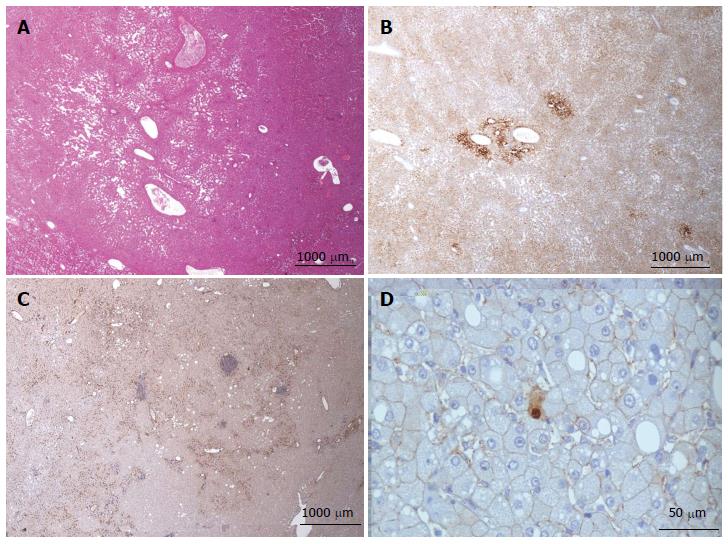
Figure 14 β-catenin activated, inflammatory hepatocellular adenoma.
A, B: Woman born in 1967. Oral contraceptives 20 years, BMI 20.0 kg/m2. By chance, discovery of one nodule 18 cm. Imaging hepatocellular adenoma (HCA). Segmentectomy IV and V 2005. A: HE: features of inflammatory HCA (IHCA): sinusoidal dilatation, thick vessels, mild inflammation. B: Glutamine synthase immunostaining is abnormal, but faint and heterogeneous with reinforcement around veins. C, D: Woman born in 1974; oral contraceptives 13 years. BMI 21.0 kg/m2. By chance, discovery of one nodule 6.5 cm. Imaging IHCA. Segmentectomy VI and VII 2009. C: Marked but not diffuse CD34 immunostaining. D: Very few tumoral hepatocytes expressed aberrant nuclear β-catenin.
- Citation: Sempoux C, Balabaud C, Bioulac-Sage P. Pictures of focal nodular hyperplasia and hepatocellular adenomas. World J Hepatol 2014; 6(8): 580-595
- URL: https://www.wjgnet.com/1948-5182/full/v6/i8/580.htm
- DOI: https://dx.doi.org/10.4254/wjh.v6.i8.580


