Copyright
©2014 Baishideng Publishing Group Inc.
World J Hepatol. Aug 27, 2014; 6(8): 580-595
Published online Aug 27, 2014. doi: 10.4254/wjh.v6.i8.580
Published online Aug 27, 2014. doi: 10.4254/wjh.v6.i8.580
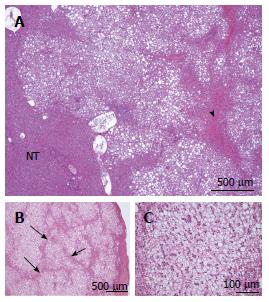
Figure 6 HNF1A mutated hepatocellular adenoma microscopic typical features.
A: Woman born in 1955; surgery in 2010 for an HCC (6 cm) developed on a non fibrotic liver. Discovery of another small nodule on the surgical specimen. No oral contraceptives; BMI 24.1 kg/m2. HE: typical aspect of HNF1A mutated hepatocellular adenoma (H-HCA): nodule with lobulated contours, made of benign hepatocytes with diffuse steatosis; some congestive areas ( arrowhead); sharp contrast with non steatotic surrounding liver. The diagnosis was confirmed by immunohistochemistry. B, C: Woman born in 1952; abdominal pain; imaging: nodule 1.8 cm, no firm diagnosis by magnetic resonance imaging. Oral contraceptives, 27 years; BMI 22.0 kg/m2. Tumorectomy in 2008. B: Nodule composed of benign, clear hepatocytes, sometimes steatotic, separated by thin strands of atrophic hepatocytes (arrow). C: Same nodule seen at higher magnification. Although clear hepatocytes are not the hallmark of H-HCA, the lobular pattern is very characteristic. It consists of steatotic or clear hepatocytes arranged in a lobular pattern separated by tumoral, atrophic, not steatotic hepatocytes. Commonly, arterioles/small arteries are seen in this space (not shown on this micrograph). The diagnosis was confirmed by immunohistochemistry.
- Citation: Sempoux C, Balabaud C, Bioulac-Sage P. Pictures of focal nodular hyperplasia and hepatocellular adenomas. World J Hepatol 2014; 6(8): 580-595
- URL: https://www.wjgnet.com/1948-5182/full/v6/i8/580.htm
- DOI: https://dx.doi.org/10.4254/wjh.v6.i8.580


