Copyright
©2014 Baishideng Publishing Group Inc.
World J Hepatol. Aug 27, 2014; 6(8): 559-569
Published online Aug 27, 2014. doi: 10.4254/wjh.v6.i8.559
Published online Aug 27, 2014. doi: 10.4254/wjh.v6.i8.559
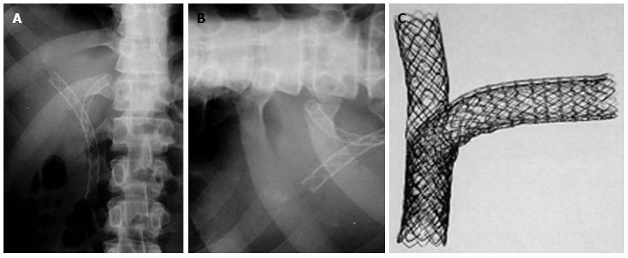
Figure 7 Percutaneous double stenting.
A: X-ray showing the Y configuration of the stent (Boston Scientific MA, United States); B: Transverse limb of a T stent (Taewoong Medial, South Korea) showing the open mesh in the center; C: Assembly of a T stent showing the vertical stent passing through the open mesh of the transverse stent.
- Citation: Goenka MK, Goenka U. Palliation: Hilar cholangiocarcinoma. World J Hepatol 2014; 6(8): 559-569
- URL: https://www.wjgnet.com/1948-5182/full/v6/i8/559.htm
- DOI: https://dx.doi.org/10.4254/wjh.v6.i8.559


