Copyright
©2014 Baishideng Publishing Group Inc.
World J Hepatol. Jul 27, 2014; 6(7): 486-495
Published online Jul 27, 2014. doi: 10.4254/wjh.v6.i7.486
Published online Jul 27, 2014. doi: 10.4254/wjh.v6.i7.486
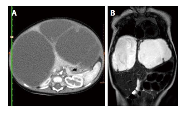
Figure 4 Abdominal computed tomography and magnetic resonance imaging.
A: Abdominal computed tomography-contrast shows enhancement of the solid component, septate, and the peripheral rim; B: Abdominal magnetic resonance imaging-contrast shows a high signal intensity on T2-weighted magnetic resonance sequences.
- Citation: Fernandez-Pineda I, Cabello-Laureano R. Differential diagnosis and management of liver tumors in infants. World J Hepatol 2014; 6(7): 486-495
- URL: https://www.wjgnet.com/1948-5182/full/v6/i7/486.htm
- DOI: https://dx.doi.org/10.4254/wjh.v6.i7.486


