Copyright
©2014 Baishideng Publishing Group Inc.
World J Hepatol. Jul 27, 2014; 6(7): 477-485
Published online Jul 27, 2014. doi: 10.4254/wjh.v6.i7.477
Published online Jul 27, 2014. doi: 10.4254/wjh.v6.i7.477
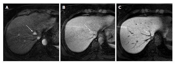
Figure 2 Magnetic resonance imaging of a small focal nodular hyperplasia.
Arterial, venous and hepatobiliary phases (A, B and C), acquired in a 44-year-old woman shows the typical enhancement of a small focal nodular hyperplasia (white arrows). The lesion is located in the fourth liver segment, between medium and left sovrahepatic vein. In hepatobiliary phase (C) the lesion is slightly hyperintense to the surrounding liver parenchyma, due to uptake of hepatospecific contrast.
- Citation: Palmucci S. Focal liver lesions detection and characterization: The advantages of gadoxetic acid-enhanced liver MRI. World J Hepatol 2014; 6(7): 477-485
- URL: https://www.wjgnet.com/1948-5182/full/v6/i7/477.htm
- DOI: https://dx.doi.org/10.4254/wjh.v6.i7.477


