Copyright
©2014 Baishideng Publishing Group Inc.
World J Hepatol. Jul 27, 2014; 6(7): 477-485
Published online Jul 27, 2014. doi: 10.4254/wjh.v6.i7.477
Published online Jul 27, 2014. doi: 10.4254/wjh.v6.i7.477
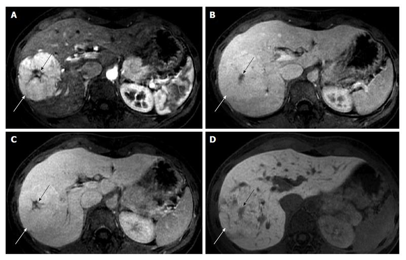
Figure 1 Typical imaging features of focal nodular hyperplasia in a 29-year-old woman.
Gadoxetic acid-enhanced magnetic resonance imaging; axial images (A-D) were obtained in dynamic phases and hepatobiliary phase. A shows a solid circumscribed mass (white arrow), lobulated in contour, with a central scar (black arrow); the lesion is hyperintense on the arterial phase (A) and persists slightly hyperintense in the portal and venous phases (B and C respectively). In hepatobiliary phase (D) the mass is slightly hyperintense or isointense to the surrounding liver. The presence of biliary canaliculi, even if not functioning, leads to retention of gadoxetic acid in comparison to the surrounding parenchyma.
- Citation: Palmucci S. Focal liver lesions detection and characterization: The advantages of gadoxetic acid-enhanced liver MRI. World J Hepatol 2014; 6(7): 477-485
- URL: https://www.wjgnet.com/1948-5182/full/v6/i7/477.htm
- DOI: https://dx.doi.org/10.4254/wjh.v6.i7.477


