Copyright
©2014 Baishideng Publishing Group Inc.
World J Hepatol. May 27, 2014; 6(5): 358-362
Published online May 27, 2014. doi: 10.4254/wjh.v6.i5.358
Published online May 27, 2014. doi: 10.4254/wjh.v6.i5.358
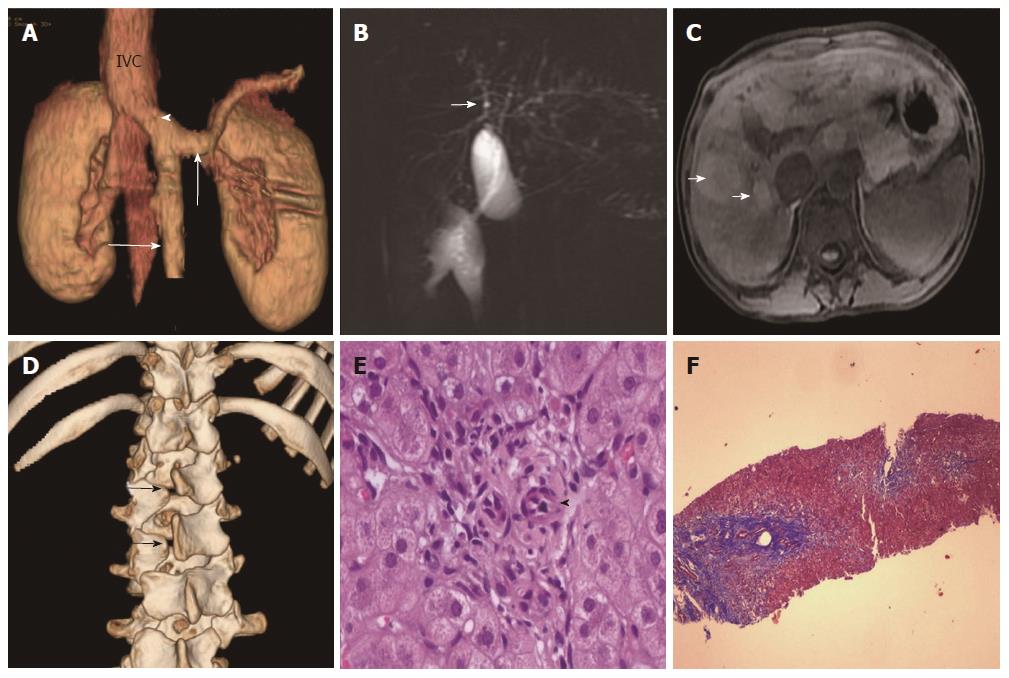
Figure 2 Congenital extrahepatic portocaval shunt (Abernethy Type 1b), multiple regenerative liver nodules and dilatation of intrahepatic biliary radicles.
A: Reconstructed 3-D images of contrast-enhanced computerized tomography (CECT) scan showing congenital extrahepatic portosystemic shunt with drainage of the portal vein (arrowhead) into the inferior vena cava-splenic and superior mesenteric veins are shown by vertical and horizontal arrows, respectively; B: Magnetic resonance cholangiopancreaticography images showing dilatation of bilobar intrahepatic biliary radicles (arrow); C: T1-weighted axial images on MR showing well defined, round, hyper-intense lesions in both liver lobes (arrows); D: Reconstructed 3-D CECT images of lumbar spine showing spina bifida at L1 and L2 levels; E, F: Liver biopsy specimen showing preserved acinar architecture; portal tract showing prominent hepatic arteriole (arrowhead) with absence of portal venule and bile ductules (E, hematoxylin and eosin stain, × 400) with periportal fibrosis (F, Masson-trichrome stain, × 40).
- Citation: Sood V, Khanna R, Alam S, Rawat D, Bhatnagar S, Rastogi A. Ductal paucity and Warkany syndrome in a patient with congenital extrahepatic portocaval shunt. World J Hepatol 2014; 6(5): 358-362
- URL: https://www.wjgnet.com/1948-5182/full/v6/i5/358.htm
- DOI: https://dx.doi.org/10.4254/wjh.v6.i5.358


