Copyright
©2014 Baishideng Publishing Group Co.
World J Hepatol. Apr 27, 2014; 6(4): 230-242
Published online Apr 27, 2014. doi: 10.4254/wjh.v6.i4.230
Published online Apr 27, 2014. doi: 10.4254/wjh.v6.i4.230
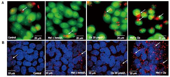
Figure 7 Autophagic detection by acridine orange staining and immunofluorescence.
Hepatocellular carcinoma (HepG2) cells were treated with 1 mmol/L melatonin and/or 20 µmol/L cisplatin for 24 h and evaluated for autophagy formation. The experiments were performed in triplicate, and representative micrographs are shown. A: Acridine orange staining showed lysosomal (red or orange) staining in the cells of all treatments. The increased acidic lysosomes in the combination treatment suggests potential lysosomal activation (scale bar = 20 μm); B: Confocal immunofluorescence micrographs of representative treated-HepG2 cells immunolabeled with the anti-LC3 antibody (scale bar = 10 μm). Melatonin and cisplatin treatment alone induced some fluorescent immunoreactivity in the cytoplasm, but the combined treatment induced intense immunoreactivity.
-
Citation: Bennukul K, Numkliang S, Leardkamolkarn V. Melatonin attenuates cisplatin-induced HepG2 cell death
via the regulation of mTOR and ERCC1 expressions. World J Hepatol 2014; 6(4): 230-242 - URL: https://www.wjgnet.com/1948-5182/full/v6/i4/230.htm
- DOI: https://dx.doi.org/10.4254/wjh.v6.i4.230


