Copyright
©2014 Baishideng Publishing Group Co.
World J Hepatol. Apr 27, 2014; 6(4): 230-242
Published online Apr 27, 2014. doi: 10.4254/wjh.v6.i4.230
Published online Apr 27, 2014. doi: 10.4254/wjh.v6.i4.230
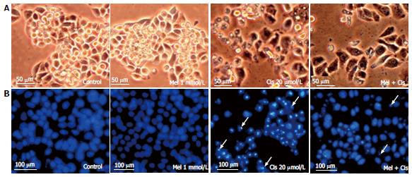
Figure 4 Histological changes.
Hepatocellular carcinoma cells were treated with melatonin (1 mmol/L) and/or cisplatin (20 μmol/L) for 48 h. The cells were stained with DAPI for fragmented DNA (apoptotic bodies) and observed under an inverted microscope. A: Morphological changes showing vacuoles in the cytoplasm of cisplatin-treated cells (scale bar = 50 μm); B: DAPI staining showing nuclear condensation and apoptotic bodies (white arrows) in cisplatin-treated cells. The combined treatment showed less apoptotic effects (scale bar = 100 μm).
-
Citation: Bennukul K, Numkliang S, Leardkamolkarn V. Melatonin attenuates cisplatin-induced HepG2 cell death
via the regulation of mTOR and ERCC1 expressions. World J Hepatol 2014; 6(4): 230-242 - URL: https://www.wjgnet.com/1948-5182/full/v6/i4/230.htm
- DOI: https://dx.doi.org/10.4254/wjh.v6.i4.230


