Copyright
©2014 Baishideng Publishing Group Co.
World J Hepatol. Mar 27, 2014; 6(3): 155-159
Published online Mar 27, 2014. doi: 10.4254/wjh.v6.i3.155
Published online Mar 27, 2014. doi: 10.4254/wjh.v6.i3.155
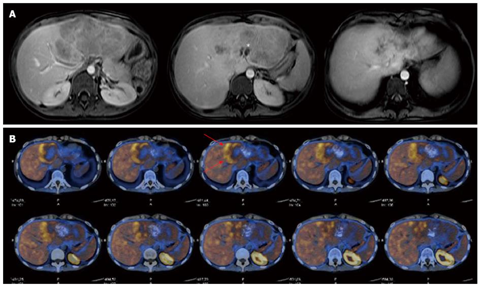
Figure 1 Radiological imaging.
A: Abdominal magnetic resonance imaging scan showing a 16 cm diameter inhomogeneous and partially calcified (asterisk) mass of the left liver; B: Total-body 11C-choline positron emission tomography showed a slight pathological uptake of the tracer in the peripheral part of the tumour (red arrows).
- Citation: Procopio F, Tommaso LD, Armenia S, Quagliuolo V, Roncalli M, Torzilli G. Nested stromal-epithelial tumour of the liver: An unusual liver entity. World J Hepatol 2014; 6(3): 155-159
- URL: https://www.wjgnet.com/1948-5182/full/v6/i3/155.htm
- DOI: https://dx.doi.org/10.4254/wjh.v6.i3.155


