Copyright
©2014 Baishideng Publishing Group Inc.
World J Hepatol. Nov 27, 2014; 6(11): 825-829
Published online Nov 27, 2014. doi: 10.4254/wjh.v6.i11.825
Published online Nov 27, 2014. doi: 10.4254/wjh.v6.i11.825
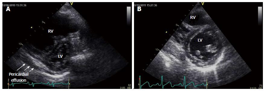
Figure 1 Transthoracic echocardiography performed before and after treatment.
A: Transthoracic echocardiography (TTE) performed before treatment. The parasternal short axis view at the basal level in diastole shows pronounced interventricular septal deviation toward the left ventricle accompanied by pericardial effusion; B: TTE performed after treatment. The parasternal short axis view at the basal level in diastole shows a decrease in the interventricular septal deviation toward the left ventricle. Pericardial effusion has disappeared. RV: Right ventricle; LV: Left ventricle.
- Citation: Yamashita Y, Tsujino I, Sato T, Yamada A, Watanabe T, Ohira H, Nishimura M. Hemodynamic effects of ambrisentan-tadalafil combination therapy on progressive portopulmonary hypertension. World J Hepatol 2014; 6(11): 825-829
- URL: https://www.wjgnet.com/1948-5182/full/v6/i11/825.htm
- DOI: https://dx.doi.org/10.4254/wjh.v6.i11.825


