Copyright
©2014 Baishideng Publishing Group Inc.
World J Hepatol. Oct 27, 2014; 6(10): 704-715
Published online Oct 27, 2014. doi: 10.4254/wjh.v6.i10.704
Published online Oct 27, 2014. doi: 10.4254/wjh.v6.i10.704
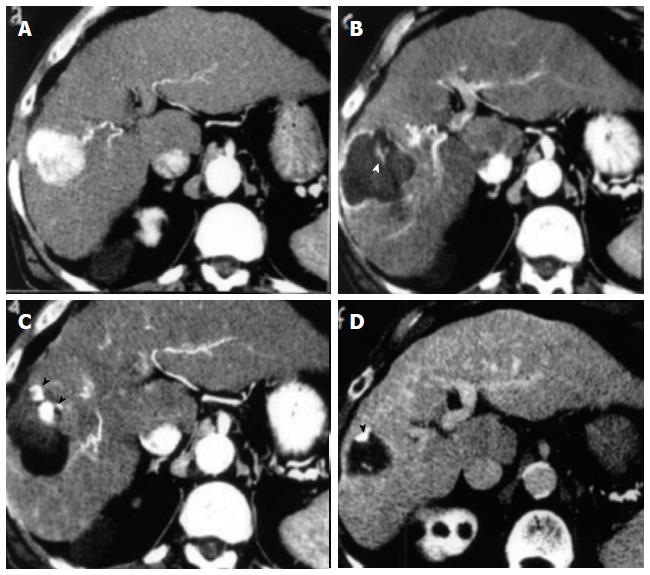
Figure 2 Representative case of complete ablation of hepatocellular carcinoma of 5 cm with combined treatment (laser ablation followed by trans-arterial-chemo-embolization).
A: Computed tomography (CT) scan before Laser ablation (LA) shows a lesion 5 cm in diameter beneath the capsule in S8 during arterial phase; B: CT scan after LA shows an area of necrosis larger than basal lesion with small viable foci (white arrowhead) within the zone of coagulation; C: CT scan shows compact retention of iodized oil in the residual viable tissue (black arrowheads) after trans-arterial-chemo-embolization (TACE) session; D: CT scan shows marked volume reduction of treated area and clear shrinkage of viable tissue (black arrowhead) 6 mo after the combined procedure.
- Citation: Costanzo GGD, Francica G, Pacella CM. Laser ablation for small hepatocellular carcinoma: State of the art and future perspectives. World J Hepatol 2014; 6(10): 704-715
- URL: https://www.wjgnet.com/1948-5182/full/v6/i10/704.htm
- DOI: https://dx.doi.org/10.4254/wjh.v6.i10.704


