Copyright
©2013 Baishideng Publishing Group Co.
World J Hepatol. Aug 27, 2013; 5(8): 417-424
Published online Aug 27, 2013. doi: 10.4254/wjh.v5.i8.417
Published online Aug 27, 2013. doi: 10.4254/wjh.v5.i8.417
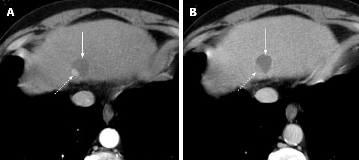
Figure 8 Incomplete hepatocellular carcinoma necrosis after percutaneous ethanol injection.
Arterial (A) and portal venous (B) computed tomography obtained 1 mo after treatment shows that approximately 10%-20% of the tumor, located in the dorsal and lateral portion of the treated, hypoattenuating area, is still viable as demonstrated by the presence of enhancement in arterial phase and hypoattenuation (“washout”) on portal venous phase (dotted arrow on A and B). The majority of tumor (solid arrow) does not show enhancement as a result of the treatment.
- Citation: Agnello F, Salvaggio G, Cabibbo G, Maida M, Lagalla R, Midiri M, Brancatelli G. Imaging appearance of treated hepatocellular carcinoma. World J Hepatol 2013; 5(8): 417-424
- URL: https://www.wjgnet.com/1948-5182/full/v5/i8/417.htm
- DOI: https://dx.doi.org/10.4254/wjh.v5.i8.417


