Copyright
©2013 Baishideng Publishing Group Co.
World J Hepatol. Aug 27, 2013; 5(8): 417-424
Published online Aug 27, 2013. doi: 10.4254/wjh.v5.i8.417
Published online Aug 27, 2013. doi: 10.4254/wjh.v5.i8.417
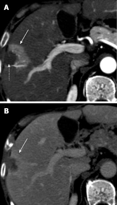
Figure 4 Perfusion alteration after radiofrequency ablation for hepatocellular carcinoma.
A: Arterial phase computed tomography (CT) obtained 1 mo after treatment shows ablated zone (dotted arrow) and a semilunar enhancing area (solid arrow) medial and anterior to ablated zone; B: Delayed phase CT shows persistent enhancement of the semilunar area (solid arrow), suggesting that the arterial enhancement is due to perfusion alteration rather than residual tumor.
- Citation: Agnello F, Salvaggio G, Cabibbo G, Maida M, Lagalla R, Midiri M, Brancatelli G. Imaging appearance of treated hepatocellular carcinoma. World J Hepatol 2013; 5(8): 417-424
- URL: https://www.wjgnet.com/1948-5182/full/v5/i8/417.htm
- DOI: https://dx.doi.org/10.4254/wjh.v5.i8.417


