Copyright
©2013 Baishideng Publishing Group Co.
World J Hepatol. Jul 27, 2013; 5(7): 404-408
Published online Jul 27, 2013. doi: 10.4254/wjh.v5.i7.404
Published online Jul 27, 2013. doi: 10.4254/wjh.v5.i7.404
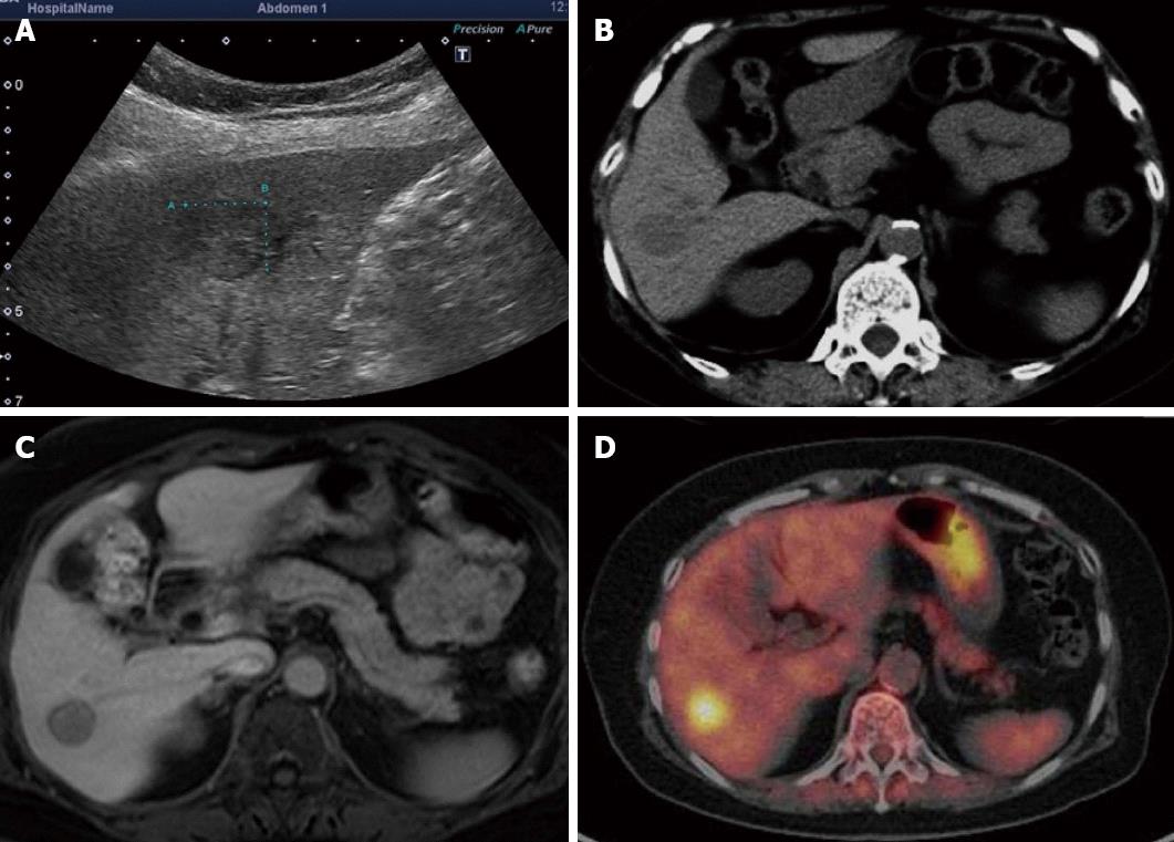
Figure 1 A mass approximately 15 mm in diameter was noted in the hepatic S6.
A: Ultrasonography; B: Computed tomography scan; C: Magnetic resonance imaging; D: Positron emission tomography.
- Citation: Miyoshi H, Mimura S, Nomura T, Tani J, Morishita A, Kobara H, Mori H, Yoneyama H, Deguchi A, Himoto T, Yamamoto N, Okano K, Suzuki Y, Masaki T. A rare case of hyaline-type Castleman disease in the liver. World J Hepatol 2013; 5(7): 404-408
- URL: https://www.wjgnet.com/1948-5182/full/v5/i7/404.htm
- DOI: https://dx.doi.org/10.4254/wjh.v5.i7.404


