Copyright
©2013 Baishideng Publishing Group Co.
World J Hepatol. Jul 27, 2013; 5(7): 398-403
Published online Jul 27, 2013. doi: 10.4254/wjh.v5.i7.398
Published online Jul 27, 2013. doi: 10.4254/wjh.v5.i7.398
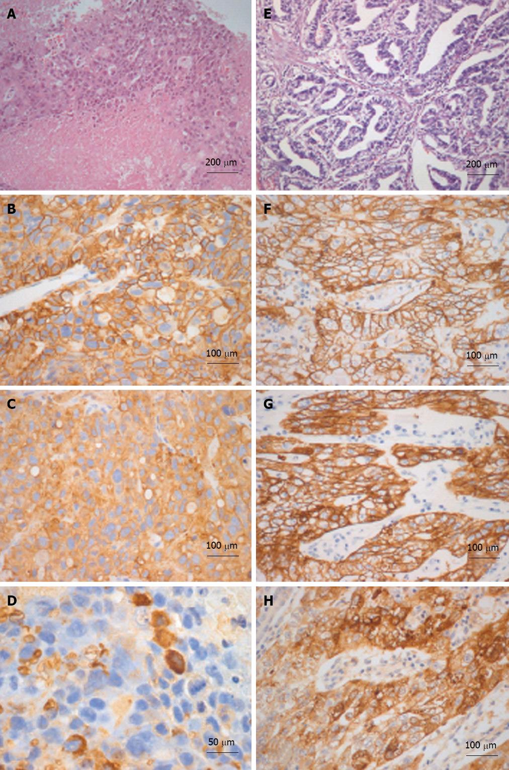
Figure 3 Histological-immunohistochemical characterizations of liver and gastric tumors are illustrated in A-D and E-H respectively.
The liver tumor is extensively necrotic (A) and neoplastic cells are immunoreactive for MOC-31 (B) and cytokeratin (CK)-18 (C). Sparse cells are immunoreactive for alpha-fetoprotein (AFP) as well (D). The gastric adenocarcinoma (E) is immunoreactive for MOC-31 (F), CK-18 (G) and AFP (H) as well. A and E: Hematoxylin and eosin.
- Citation: Cardinale V, De Filippis G, Corsi A, La Penna A, Rossi M, Catalano C, Bianco P, De Santis A, Alvaro D. An isolate alpha-fetoprotein producing gastric cancer liver metastasis emerged in a patient previously affected by radiation induced liver disease. World J Hepatol 2013; 5(7): 398-403
- URL: https://www.wjgnet.com/1948-5182/full/v5/i7/398.htm
- DOI: https://dx.doi.org/10.4254/wjh.v5.i7.398


