Copyright
©2013 Baishideng Publishing Group Co.
World J Hepatol. Jul 27, 2013; 5(7): 398-403
Published online Jul 27, 2013. doi: 10.4254/wjh.v5.i7.398
Published online Jul 27, 2013. doi: 10.4254/wjh.v5.i7.398
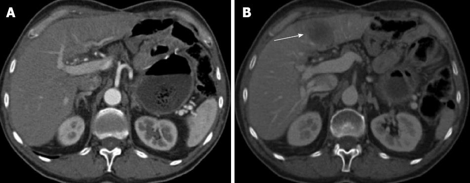
Figure 2 Axial computed tomography scan after iv iodinated contrast medium administration in the arterial hepatic phase (A) and portal phase (B).
A: The computed tomography study does not show focal lesions in the liver parenchyma (Jul 2008); B: A focal nodular hypo attenuated lesion is present affecting the left hepatic lobe (arrow); it presents with regular margins and appears mildly vascularised (Nov 2008).
- Citation: Cardinale V, De Filippis G, Corsi A, La Penna A, Rossi M, Catalano C, Bianco P, De Santis A, Alvaro D. An isolate alpha-fetoprotein producing gastric cancer liver metastasis emerged in a patient previously affected by radiation induced liver disease. World J Hepatol 2013; 5(7): 398-403
- URL: https://www.wjgnet.com/1948-5182/full/v5/i7/398.htm
- DOI: https://dx.doi.org/10.4254/wjh.v5.i7.398


