Copyright
©2013 Baishideng Publishing Group Co.
World J Hepatol. May 27, 2013; 5(5): 288-291
Published online May 27, 2013. doi: 10.4254/wjh.v5.i5.288
Published online May 27, 2013. doi: 10.4254/wjh.v5.i5.288
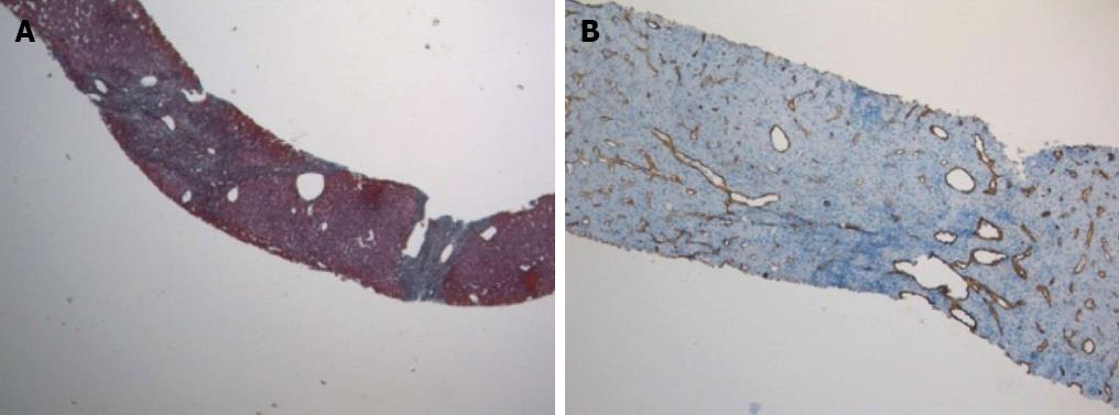
Figure 1 Focal nodular hyperplasia.
A: Fibrous septa (green) giving a nodular appearance to liver parenchyma. Masson’s trichrome staining; original magnification × 40; B: Diffuse CD34 positive immunostaining in the sinusoids; original magnification × 100.
- Citation: Macaluso FS, Maida M, Alessi N, Cabibbo G, Cabibi D. Primary biliary cirrhosis and hereditary hemorrhagic telangiectasia: When two rare diseases coexist. World J Hepatol 2013; 5(5): 288-291
- URL: https://www.wjgnet.com/1948-5182/full/v5/i5/288.htm
- DOI: https://dx.doi.org/10.4254/wjh.v5.i5.288


