Copyright
©2013 Baishideng Publishing Group Co.
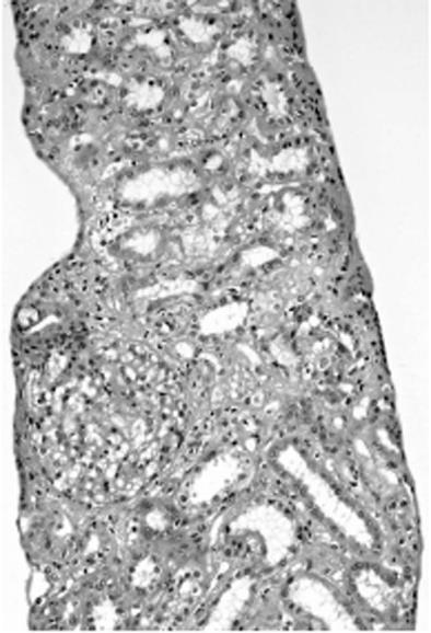
Figure 2 Light microscopic findings of the renal tissue showing dilated renal tubules, edema and inflammatory infiltrate in the interstitium (hematoxylin and eosin stain, ×100).
- Citation: Kishi T, Ikeda Y, Takashima T, Rikitake S, Miyazono M, Aoki S, Sakemi T, Mizuta T, Fujimoto K. Acute renal failure associated with acute non-fulminant hepatitis B. World J Hepatol 2013; 5(2): 82-85
- URL: https://www.wjgnet.com/1948-5182/full/v5/i2/82.htm
- DOI: https://dx.doi.org/10.4254/wjh.v5.i2.82


