Copyright
©2012 Baishideng Publishing Group Co.
World J Hepatol. Jul 27, 2012; 4(7): 199-208
Published online Jul 27, 2012. doi: 10.4254/wjh.v4.i7.199
Published online Jul 27, 2012. doi: 10.4254/wjh.v4.i7.199
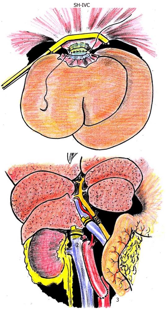
Figure 7 Orthotopic liver transplant in the rat.
The first anastomosis that is carried out in the orthotopic transplant of the rat's liver is end-to-end between the suprahepatic inferior vena cava of the donor and recipient is represented on the top. The suture of the posterior semi circumferences of both vessels is intraluminal and from left to right of the animal. Next, the extraluminal suture is made on the anterior semi circumferences in the same way. Before finishing this anastomosis, saline is perfused inside to eliminate air that can cause a gas embolism when revascularizing the transplanted liver. The end-to-end portal anastomosis is made second, using the cuff technique. It is convenient to perfuse the liver through the portal route before the anastomosis is completed to eliminate potassium and acid catabolites, which accumulated during hypothermic preservation, from its circulation. Once proceeding with portal revascularization of the liver, the end-to-end anastomosis of the infrahepatic inferior vena cava of the donor and recipient is performed using the cuff technique. Then, arterial revascularization using the donor aorta, in continuity with the celiac trunk and hepatic artery, is made. End-to-side anastomosis is performed with the infrarenal abdominal aorta of the recipient. Lastly, the choledococholedochostomy is performed (bottom). 1, portal vein anastomosis; 2, infrahepatic inferior vena cava anastomosis; 3, arterial anastomosis; 4, choledococholedochostomy.
- Citation: Aller MA, Arias N, Prieto I, Agudo S, Gilsanz C, Lorente L, Arias JL, Arias J. A half century (1961-2011) of applying microsurgery to experimental liver research. World J Hepatol 2012; 4(7): 199-208
- URL: https://www.wjgnet.com/1948-5182/full/v4/i7/199.htm
- DOI: https://dx.doi.org/10.4254/wjh.v4.i7.199


