Copyright
©2012 Baishideng Publishing Group Co.
World J Hepatol. Jun 27, 2012; 4(6): 191-195
Published online Jun 27, 2012. doi: 10.4254/wjh.v4.i6.191
Published online Jun 27, 2012. doi: 10.4254/wjh.v4.i6.191
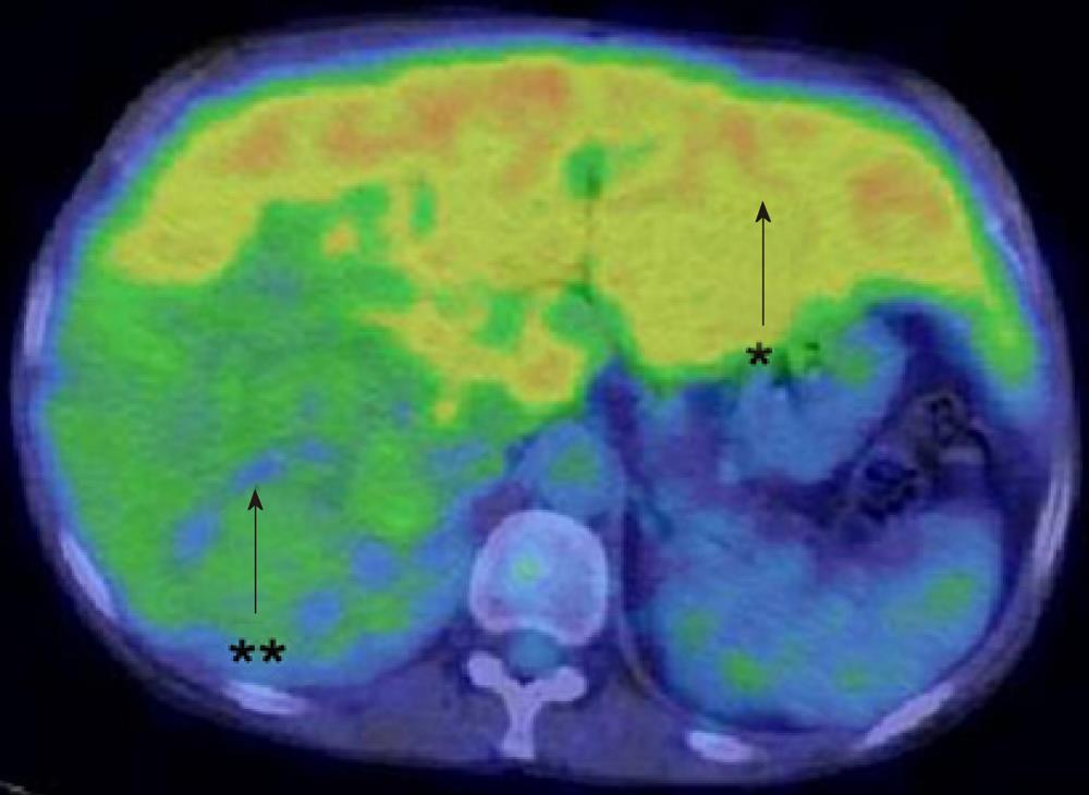
Figure 5 Fluorodeoxyglucose-positron emission tomography/computer tomography.
High levels of FDG accumulation with a SUVmax of 4.2 in the non-tumorous liver (arrow, star) and relatively low uptake of FDG with a SUVmax of 2.7 in the tumor (arrow, double star). FDG: Fluorodeoxyglucose-positron emission tomography.
- Citation: Mikuriya Y, Oshita A, Tashiro H, Amano H, Kobayashi T, Arihiro K, Ohdan H. Hepatocellular carcinoma and focal nodular hyperplasia of the liver in a glycogen storage disease patient. World J Hepatol 2012; 4(6): 191-195
- URL: https://www.wjgnet.com/1948-5182/full/v4/i6/191.htm
- DOI: https://dx.doi.org/10.4254/wjh.v4.i6.191


