Copyright
©2012 Baishideng Publishing Group Co.
World J Hepatol. Nov 27, 2012; 4(11): 322-326
Published online Nov 27, 2012. doi: 10.4254/wjh.v4.i11.322
Published online Nov 27, 2012. doi: 10.4254/wjh.v4.i11.322
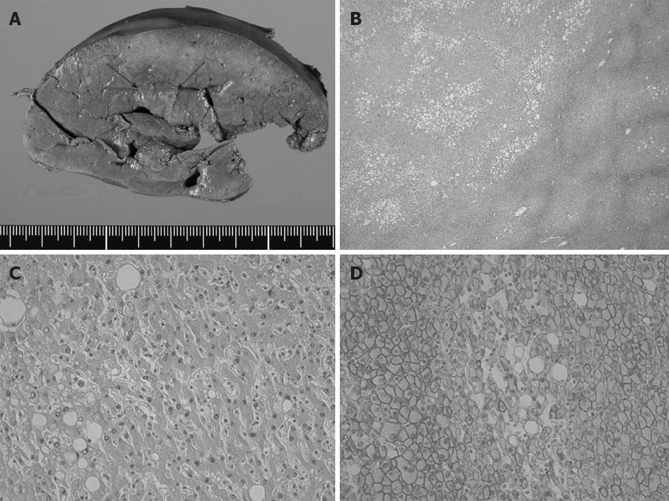
Figure 3 Pathological findings of the liver tumor.
A: Cut surface of formalin-fixed liver specimen. The tumor was unencapsulated and its border was ill-defined (arrow); B, C: Microscopically, the tumor consisted of low-grade atypical hepatocytes, without cellular mitosis or changes in cellular density and structures. Fatty deposition in the tumor cells was ubiquitously remarkable. Neither biliary structures nor portal triads were present within the tumor (hematoxylin-eosin stain, original magnification ×40 in B and ×200 in C); D: β-catenin was immunohistochemically detected on the cytomembrane. Neither aberrant nuclear nor cytoplasmic accumulations were found (original magnification ×100).
- Citation: Inaba K, Sakaguchi T, Kurachi K, Mori H, Tao H, Nakamura T, Takehara Y, Baba S, Maekawa M, Sugimura H, Konno H. Hepatocellular adenoma associated with familial adenomatous polyposis coli. World J Hepatol 2012; 4(11): 322-326
- URL: https://www.wjgnet.com/1948-5182/full/v4/i11/322.htm
- DOI: https://dx.doi.org/10.4254/wjh.v4.i11.322


