Copyright
©2012 Baishideng Publishing Group Co.
World J Hepatol. Oct 27, 2012; 4(10): 274-283
Published online Oct 27, 2012. doi: 10.4254/wjh.v4.i10.274
Published online Oct 27, 2012. doi: 10.4254/wjh.v4.i10.274
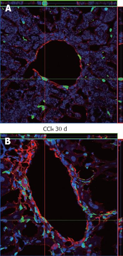
Figure 9 Immunostaining analysis of α-smooth muscle actin in liver samples from chimeric animals by confocal microscopy.
A: α-smooth muscle actin (α-SMA)+ cells in the perivascular region were observed where green fluorescent protein (GFP)+ cells could also be seen, but the two cell types do not co-localize, as can be seen by the orientation of the red and green bars; B: An increase in reactivity against α-SMA around the perivascular region was observed, also with augmentation of GFP+ cells, but again these cell types do not co-localize. In both images the nuclei were stained with TO-PRO3 (blue).
- Citation: Paredes BD, Faccioli LAP, Quintanilha LF, Asensi KD, Valle CZD, Canary PC, Takiya CM, Carvalho ACC, Goldenberg RCDS. Bone marrow progenitor cells do not contribute to liver fibrogenic cells. World J Hepatol 2012; 4(10): 274-283
- URL: https://www.wjgnet.com/1948-5182/full/v4/i10/274.htm
- DOI: https://dx.doi.org/10.4254/wjh.v4.i10.274


