Copyright
©2011 Baishideng Publishing Group Co.
World J Hepatol. Jul 27, 2011; 3(7): 184-197
Published online Jul 27, 2011. doi: 10.4254/wjh.v3.i7.184
Published online Jul 27, 2011. doi: 10.4254/wjh.v3.i7.184
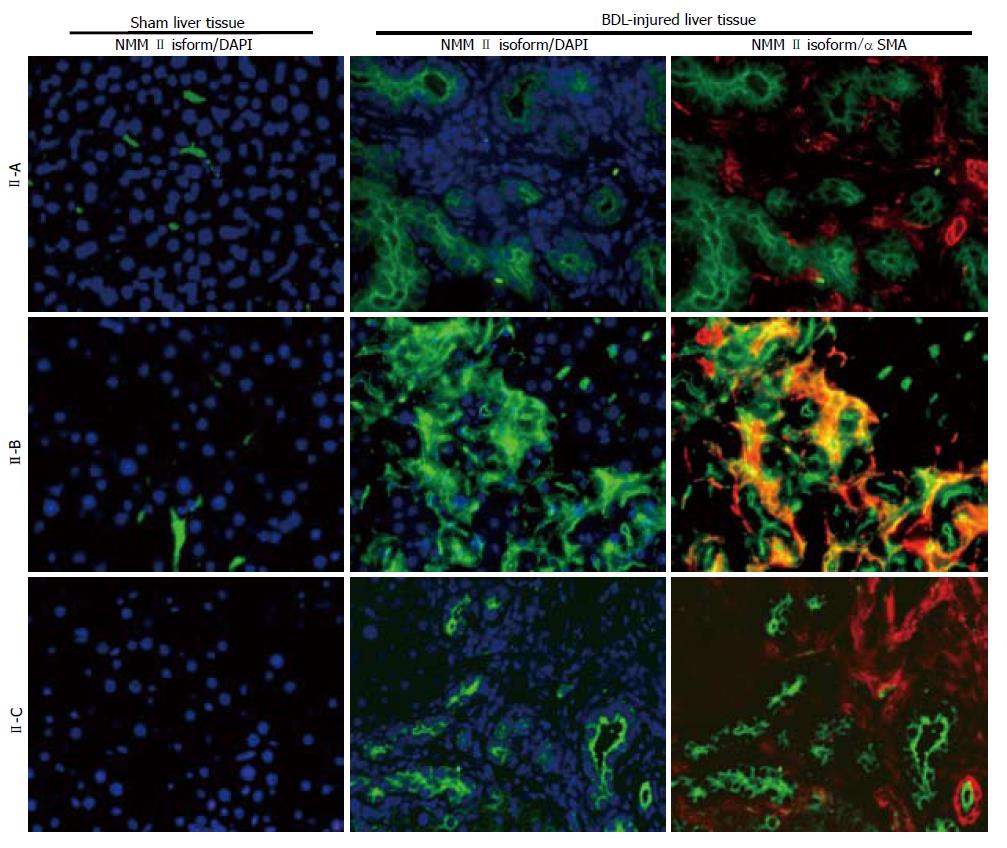
Figure 3 Immunohistochemical analysis of nonmuscle myosin II-A, II-B and II-C in normal and injured liver sections.
Specific nonmuscle myosin (NMM) II immunoreactivity of representative fields is shown in green; αactin smooth muscle, as a marker of hepatic stellate cell activation is shown in red and cell nuclei are stained blue (DAPI). Images of NMM II isoforms, F-actin, and DAPI were taken separately at identical exposures and color channels were merged using IMAGE-PRO software (200 ×).
- Citation: Moore CC, Lakner AM, Yengo CM, Schrum LW. Nonmuscle myosin II regulates migration but not contraction in rat hepatic stellate cells. World J Hepatol 2011; 3(7): 184-197
- URL: https://www.wjgnet.com/1948-5182/full/v3/i7/184.htm
- DOI: https://dx.doi.org/10.4254/wjh.v3.i7.184


