Copyright
©2011 Baishideng Publishing Group Co.
World J Hepatology. Oct 27, 2011; 3(10): 271-274
Published online Oct 27, 2011. doi: 10.4254/wjh.v3.i10.271
Published online Oct 27, 2011. doi: 10.4254/wjh.v3.i10.271
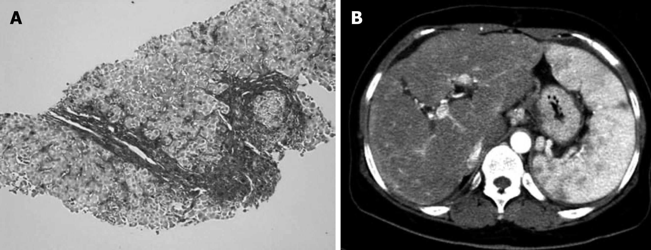
Figure 3 Second histological examination of the liver, and the image enhanced computed tomogram.
A: The second biopsy revealed pseudo-lobular dense fibrosis with moderate infiltrating cells; B: The images of the enhanced computed tomography (CT) were significantly altered, too. The enhanced CT at the second biopsy showed a marked splenomegaly, and the surface of the liver was irregular, indicating portal hypertension and liver cirrhosis, respectively.
- Citation: Yoshiji H, Kitagawa K, Noguchi R, Uemura M, Ikenaka Y, Aihara Y, Nakanishi K, Shirai Y, Morioka C, Fukui H. A histologically proven case of progressive liver sarcoidosis with variceal rupture. World J Hepatology 2011; 3(10): 271-274
- URL: https://www.wjgnet.com/1948-5182/full/v3/i10/271.htm
- DOI: https://dx.doi.org/10.4254/wjh.v3.i10.271


