Copyright
©2011 Baishideng Publishing Group Co.
World J Hepatology. Oct 27, 2011; 3(10): 271-274
Published online Oct 27, 2011. doi: 10.4254/wjh.v3.i10.271
Published online Oct 27, 2011. doi: 10.4254/wjh.v3.i10.271
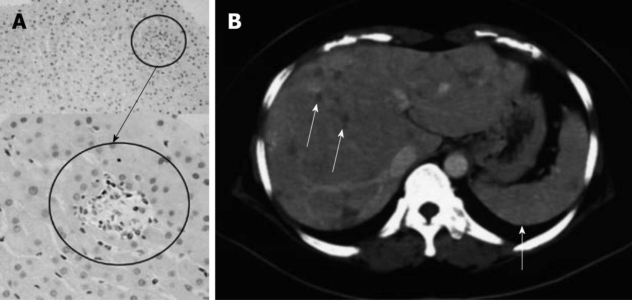
Figure 2 First histological examination of the liver, and the image of enhanced computed tomogram.
A: The first liver biopsy showed non-caseating granulomas in the liver without fibrosis development. Aggregates of epithelioid histiocytes and Langhans-type giant cells were observed surrounded by lymphocytes. The original magnifications are × 40 and × 200, respectively; B: Enhanced computed tomogram showing multiple low-attenuation areas up to 10 mm in diameter, indicating multiple granulomas in the liver (white arrows). There was no splenomegaly at this time.
- Citation: Yoshiji H, Kitagawa K, Noguchi R, Uemura M, Ikenaka Y, Aihara Y, Nakanishi K, Shirai Y, Morioka C, Fukui H. A histologically proven case of progressive liver sarcoidosis with variceal rupture. World J Hepatology 2011; 3(10): 271-274
- URL: https://www.wjgnet.com/1948-5182/full/v3/i10/271.htm
- DOI: https://dx.doi.org/10.4254/wjh.v3.i10.271


