Copyright
©2010 Baishideng Publishing Group Co.
World J Hepatol. Aug 27, 2010; 2(8): 311-317
Published online Aug 27, 2010. doi: 10.4254/wjh.v2.i8.311
Published online Aug 27, 2010. doi: 10.4254/wjh.v2.i8.311
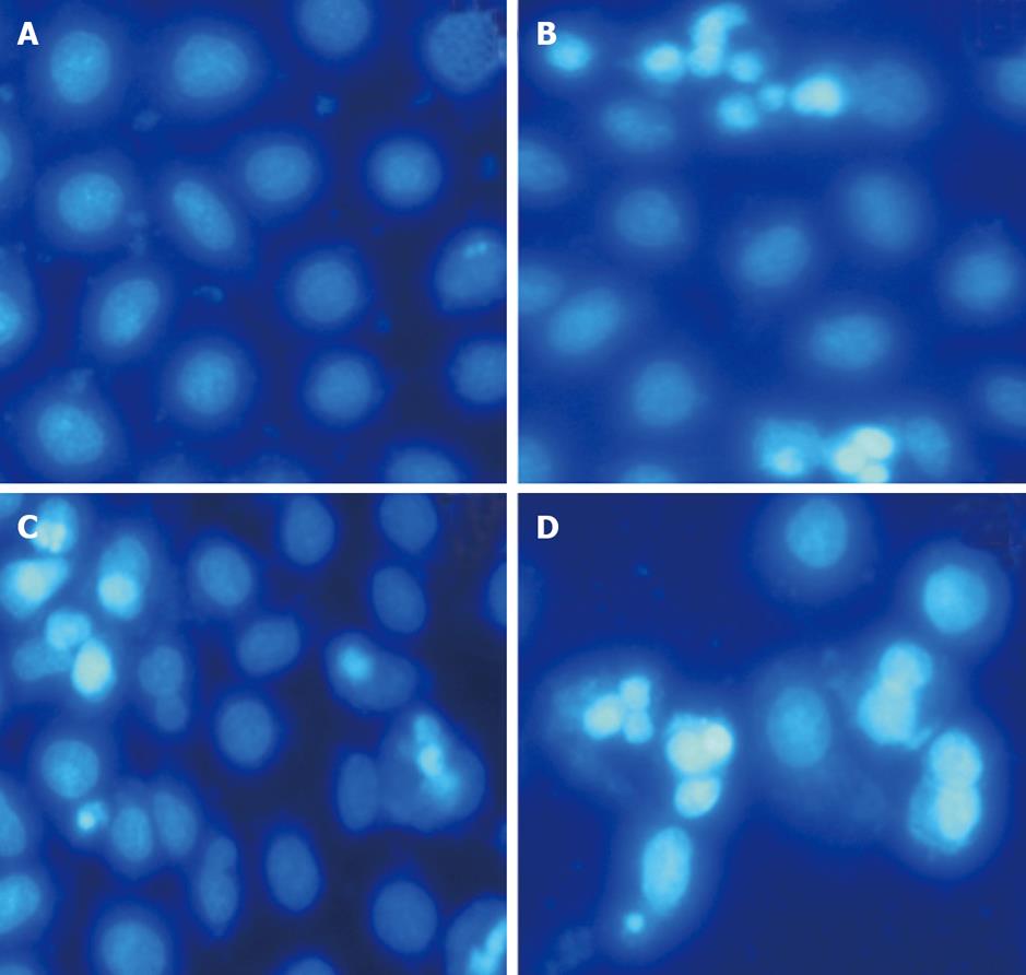
Figure 3 Morphological changes in the apoptotic cells after Hoechst 33258 staining (×200).
A: Untreated HepG-2 cells; B: HepG-2 cells treated with 100 μg/mL mofetil (MMF) for 48 h; C: HepG-2 cells treated with 1 μg/mL adriamycin (ADM) for 48 h; D: HepG-2 cells treated with MMF + ADM for 48 h.
- Citation: Chu YK, Liu Y, Yin JK, Wang N, Cai L, Lu JG. Effect of mycophenolate mofetil plus adriamycin on HepG-2 cells. World J Hepatol 2010; 2(8): 311-317
- URL: https://www.wjgnet.com/1948-5182/full/v2/i8/311.htm
- DOI: https://dx.doi.org/10.4254/wjh.v2.i8.311


