Copyright
©2010 Baishideng.
World J Hepatol. May 27, 2010; 2(5): 192-197
Published online May 27, 2010. doi: 10.4254/wjh.v2.i5.192
Published online May 27, 2010. doi: 10.4254/wjh.v2.i5.192
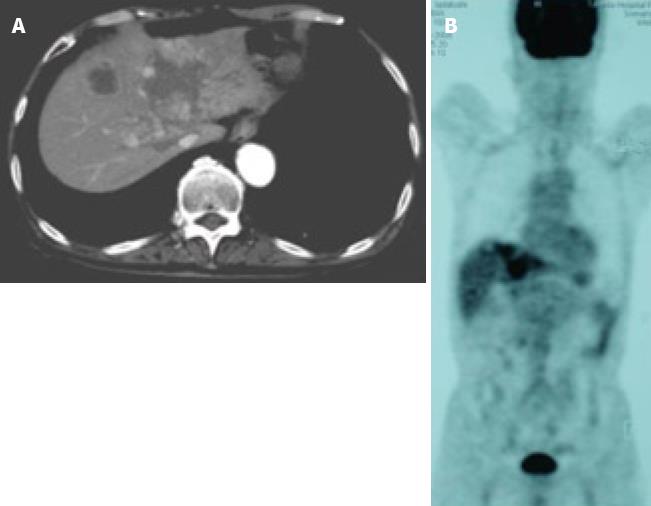
Figure 4 Images after the end of 7 cycles of chemotherapy.
A: CE-CT showed a reduction in the size of a huge tumor seen in the medial segment; B: PET-CT revealed abnormal uptake only in the tumors in the medial segment, and no uptake was observed in tumors from the right lobe and the LNs in the liver hilus and para-aorta.
- Citation: Nishimura M. A successful treatment by hepatic arterial infusion therapy for advanced, unresectable biliary tract cancer. World J Hepatol 2010; 2(5): 192-197
- URL: https://www.wjgnet.com/1948-5182/full/v2/i5/192.htm
- DOI: https://dx.doi.org/10.4254/wjh.v2.i5.192


