Copyright
©2010 Baishideng.
World J Hepatol. May 27, 2010; 2(5): 192-197
Published online May 27, 2010. doi: 10.4254/wjh.v2.i5.192
Published online May 27, 2010. doi: 10.4254/wjh.v2.i5.192
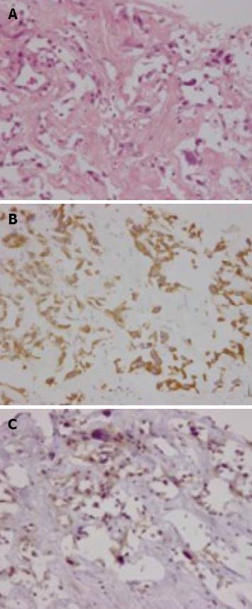
Figure 2 Biopsied specimens from one of the hepatic tumors.
A: A moderately-differentiated adenocarcinoma with wide-spread necrosis, which were not accompanied by hepatic cell components (HE × 200); B: Positive immunostaining for cytokeratin 7 (× 200) in the adenocarcinoma cells; C: Immuno-positive AFP (× 200) in the adenocarcinoma cells.
- Citation: Nishimura M. A successful treatment by hepatic arterial infusion therapy for advanced, unresectable biliary tract cancer. World J Hepatol 2010; 2(5): 192-197
- URL: https://www.wjgnet.com/1948-5182/full/v2/i5/192.htm
- DOI: https://dx.doi.org/10.4254/wjh.v2.i5.192


