Copyright
©2010 Baishideng.
World J Hepatol. May 27, 2010; 2(5): 192-197
Published online May 27, 2010. doi: 10.4254/wjh.v2.i5.192
Published online May 27, 2010. doi: 10.4254/wjh.v2.i5.192
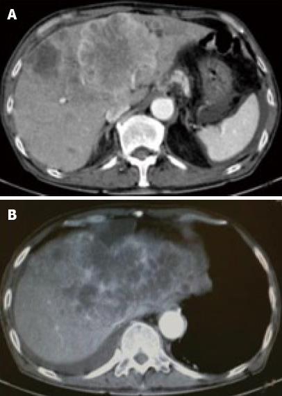
Figure 1 Contrast-enhanced (CE) computed tomography of the abdomen revealed a huge tumor, measuring 11 cm in diameter, which presented with ring enhancement in the arterial phase and central necrosis.
A: In the medial segment; B: Multiple small-sized tumors mainly located in the anterior and medial segments of the liver.
- Citation: Nishimura M. A successful treatment by hepatic arterial infusion therapy for advanced, unresectable biliary tract cancer. World J Hepatol 2010; 2(5): 192-197
- URL: https://www.wjgnet.com/1948-5182/full/v2/i5/192.htm
- DOI: https://dx.doi.org/10.4254/wjh.v2.i5.192


