Copyright
©2010 Baishideng.
World J Hepatol. May 27, 2010; 2(5): 175-179
Published online May 27, 2010. doi: 10.4254/wjh.v2.i5.175
Published online May 27, 2010. doi: 10.4254/wjh.v2.i5.175
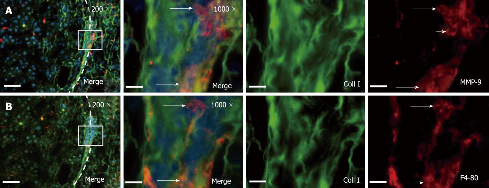
Figure 2 Conglomerates of tumor associated macrophages express MMP-9 within the HCC tumor capsule.
A: Immunofluorescence co-staining of collagen type I (green) and MMP-9 (red) displayed clusters of MMP-9 expressing cells in the tumor capsule. B: After photobleaching of the red fluorescence dye, macrophage marker F4/80 was visualized. The long arrows indicate MMP-9 positive macrophages. The shot arrow indicates an MMP-9 expressing cell which is negative for F4/80. DAPI was used for nucleus staining. The left panels provide a section overview with 200-fold magnification and bars represent 100 μm (left side of the panel: Tumor; dashed line: Tumor capsule; box: This is enlarged to 1000 × magnification in the corresponding panel, bars represent 20 μm). Original green and red channel micrographs (1000 ×) were shown. Images were derived by conventional wide field fluorescence microscopy. Representative digital overlays are shown.
- Citation: Roderfeld M, Rath T, Lammert F, Dierkes C, Graf J, Roeb E. Innovative immunohistochemistry identifies MMP-9 expressing macrophages at the invasive front of murine HCC. World J Hepatol 2010; 2(5): 175-179
- URL: https://www.wjgnet.com/1948-5182/full/v2/i5/175.htm
- DOI: https://dx.doi.org/10.4254/wjh.v2.i5.175


