Copyright
©2010 Baishideng.
World J Hepatol. Apr 27, 2010; 2(4): 167-170
Published online Apr 27, 2010. doi: 10.4254/wjh.v2.i4.167
Published online Apr 27, 2010. doi: 10.4254/wjh.v2.i4.167
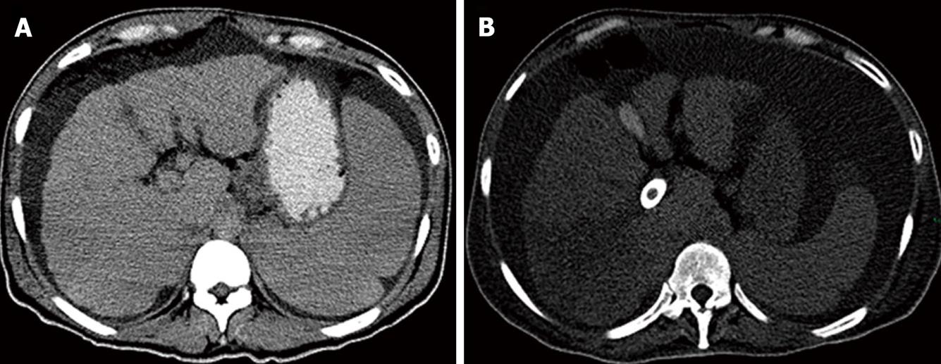
Figure 2 Abdominal computed tomography evaluation.
A: Abdominal CT scan one year prior to TIPS placement showing a small nodular liver with ascites and splenomegaly; B: Abdominal CT scan one day after TIPS placement showing a hypodense triangular-shaped heterogeneous area in the right posterior segments of the liver, suggestive of hepatic ischemia; note how the spleen size is significantly reduced.
- Citation: López-Méndez E, Zamora-Valdés D, Díaz-Zamudio M, Fernández-Díaz OF, Ávila L. Liver failure after an uncovered TIPS procedure associated with hepatic infarction. World J Hepatol 2010; 2(4): 167-170
- URL: https://www.wjgnet.com/1948-5182/full/v2/i4/167.htm
- DOI: https://dx.doi.org/10.4254/wjh.v2.i4.167


