Copyright
©2010 Baishideng.
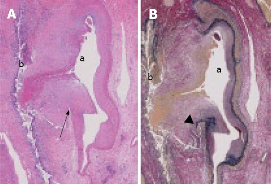
Figure 3 Histological findings revealing that the intrahepatic artery had ruptured into the intrahepatic bile duct (arrows).
a: artery; b: bile duct. The inner membrane, elastic laminae, and tunica media of the artery could not be detected at the side of rupture (arrowhead). A: HE stain, × 10; B: Elastica van Gieson stain, × 10.
- Citation: Takeda K, Tanaka K, Endo I, Togo S, Shimada H. Two-stage treatment of an unusual haemobilia caused by intrahepatic pseudoaneurysm. World J Hepatol 2010; 2(1): 52-54
- URL: https://www.wjgnet.com/1948-5182/full/v2/i1/52.htm
- DOI: https://dx.doi.org/10.4254/wjh.v2.i1.52


