Copyright
©2010 Baishideng.
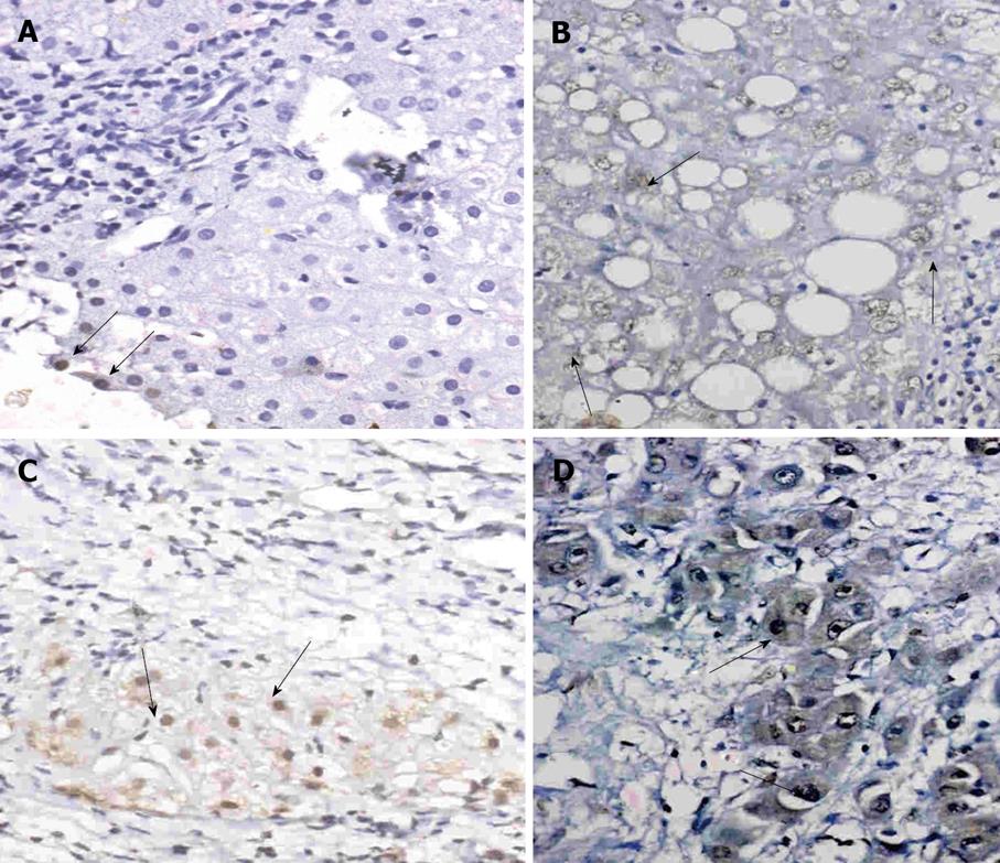
Figure 1 Immunohistochemical staining for Cyclin D1 in liver sections of patients from the control group as well as HCV-infected patients and HCV-related HCC patients.
A: Liver section from a control case showing mild nuclear staining for Cyclin D1 (arrows) (Immunoperoxidase × 40); B: Liver section from a case of CHC showing mild nuclear expression of Cyclin D1 (arrows). The portal tract is completely free from immunostaining (Immunoperoxidase × 40); C: Liver section from a case of LC showing moderate number of positively stained hepatocytes in a cirrhotic nodule (arrows) (Immunoperoxidase × 40); D: Liver section from a case of poorly-differentiated HCC showing marked nuclear expression of Cyclin D1 (arrows) (Immunoperoxidase × 40).
- Citation: Bassiouny AEE, Nosseir MM, Zoheiry MK, Ameen NA, Abdel-Hadi AM, Ibrahim IM, Zada S, El-Deen AHS, El-Bassiouni NE. Differential expression of cell cycle regulators in HCV-infection and related hepatocellular carcinoma. World J Hepatol 2010; 2(1): 32-41
- URL: https://www.wjgnet.com/1948-5182/full/v2/i1/32.htm
- DOI: https://dx.doi.org/10.4254/wjh.v2.i1.32


