Copyright
©The Author(s) 2025.
World J Hepatol. Feb 27, 2025; 17(2): 101691
Published online Feb 27, 2025. doi: 10.4254/wjh.v17.i2.101691
Published online Feb 27, 2025. doi: 10.4254/wjh.v17.i2.101691
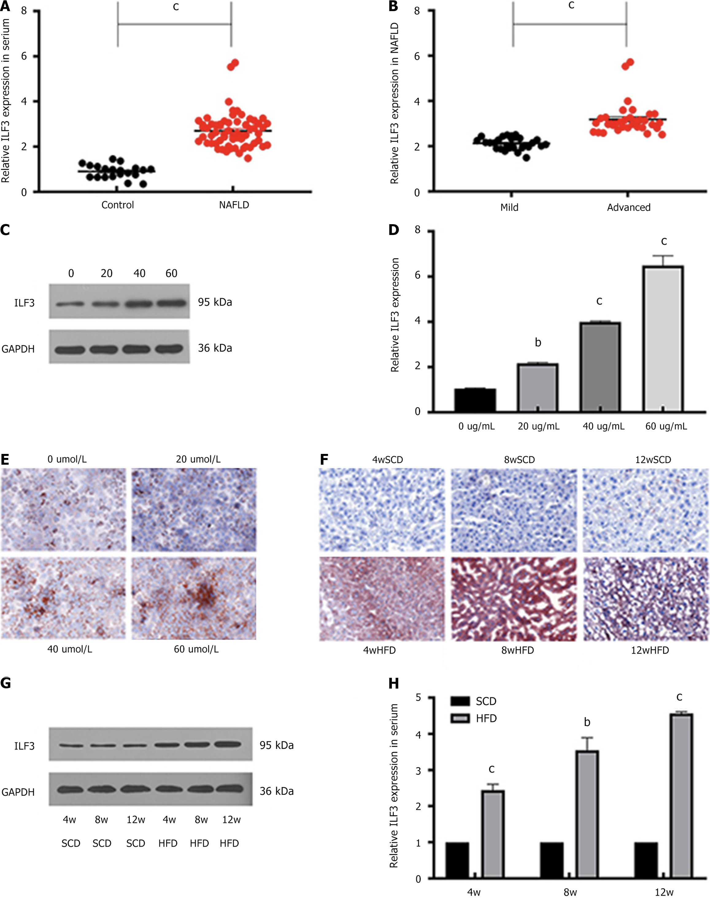
Figure 1 Elevated ILF3 expression in non-alcoholic fatty liver disease patients and models.
A: ILF3 expression in serum of non-alcoholic fatty liver disease (NAFLD) patients; B: Serum ILF3 expression according to NAFLD severity; C and D: Elevated ILF3 expression in rising Oleic acid mixture (OA) concentration treated in HepG2 cells; E and F: Oil Red O staining showing increased lipid deposition in OA-treated HepG2 cells and mice liver tissues; G and H: Elevated ILF3 expression in NAFLD mice model detected by qPCR and Western blotting. All data are presented as the mean ± SD. bP < 0.01; cP < 0.001; NAFLD: Non-alcoholic fatty liver disease; SCD: Standard chow diet; HFD: High-fat diet.
- Citation: Zhan T, Liu JX, Huang M, Chen MT, Tian XR, Yang XL, Tan J, Zou YL, Han Z, Chen W, Tian X, Huang XD. ILF3 inhibits p-AMPK expression to drive non-alcoholic fatty liver disease progression. World J Hepatol 2025; 17(2): 101691
- URL: https://www.wjgnet.com/1948-5182/full/v17/i2/101691.htm
- DOI: https://dx.doi.org/10.4254/wjh.v17.i2.101691


