Copyright
©The Author(s) 2025.
World J Hepatol. Feb 27, 2025; 17(2): 100923
Published online Feb 27, 2025. doi: 10.4254/wjh.v17.i2.100923
Published online Feb 27, 2025. doi: 10.4254/wjh.v17.i2.100923
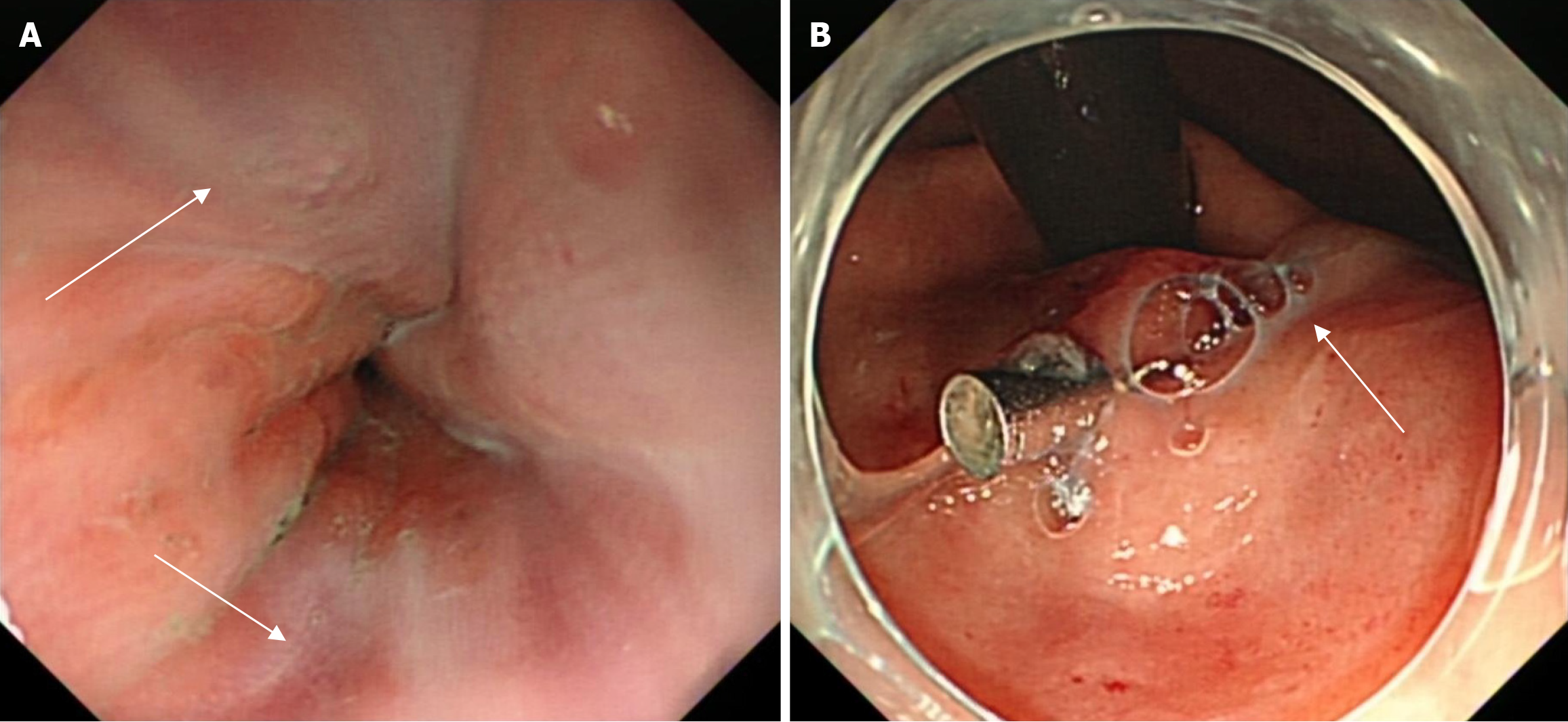
Figure 1 Endoscopic presentation of esophageal varices.
Treatment: 1 titanium clip was inserted to restrict varicose vein flow at the cardia. The varicose vein was embolized using lauromacrogol injection + hydroxyl acrylate + lauromacrogol injection, and the varicose vein in the esophagus was perfused with polyglutethimide. A: Several varicose veins are observed 30 cm from the incisors (white arrow); B: One varicose vein is observed on the side of the cardia’s lesser curvature (white arrow).
- Citation: Liu XC, Yan HH, Wei W, Du Q. Idiopathic portal hypertension misdiagnosed as hepatitis B cirrhosis: A case report and review of the literature. World J Hepatol 2025; 17(2): 100923
- URL: https://www.wjgnet.com/1948-5182/full/v17/i2/100923.htm
- DOI: https://dx.doi.org/10.4254/wjh.v17.i2.100923


