Copyright
©The Author(s) 2025.
World J Hepatol. Jan 27, 2025; 17(1): 101664
Published online Jan 27, 2025. doi: 10.4254/wjh.v17.i1.101664
Published online Jan 27, 2025. doi: 10.4254/wjh.v17.i1.101664
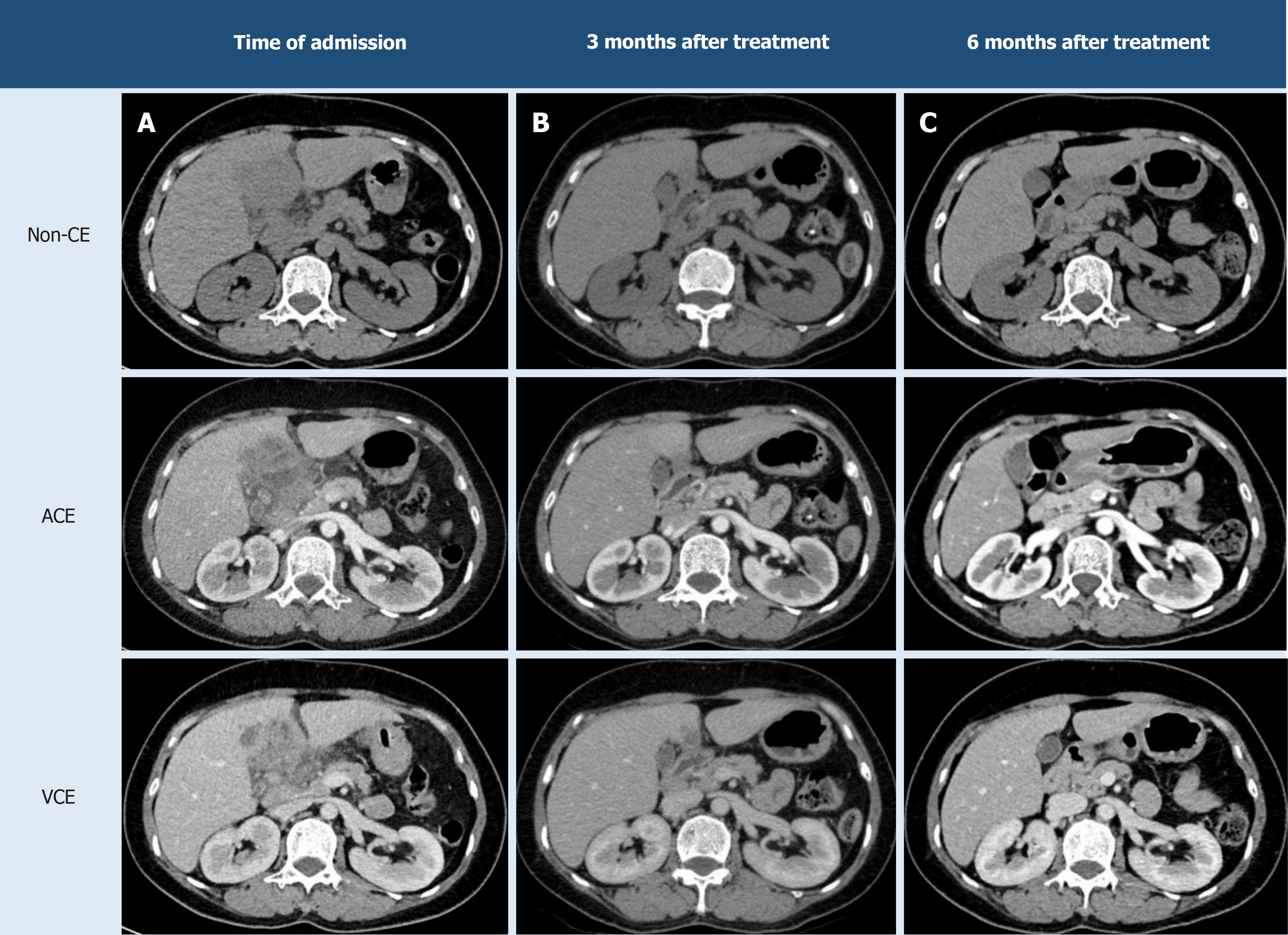
Figure 4 Morphological changes of the hepatic hilum lesion on computed tomography scans post-treatment.
A: The hepatic lesion in segment IV was initially suspected to be a biliary tumor; B: Three months after a single dose of triclabendazole, the lesion showed no features of a biliary neoplasm. Only a few small, low-attenuation lesions remained in both liver lobes, with no localization to the hepatic hilum; C: By 6 months post-treatment, the original lesion was nearly resolved, and the gallbladder showed no signs of tumor invasion. ACE: Arterial phase contrast enhanced; Non-CE: Non-contrast enhanced; VCE: Venous phase contrast enhanced.
- Citation: Le KL, Tran MQ, Pham TN, Duong NNQ, Dinh TT, Le NK. Hepatic eosinophilic pseudotumor due to Fasciola hepatica infection mimicking intrahepatic cholangiocarcinoma: A case report. World J Hepatol 2025; 17(1): 101664
- URL: https://www.wjgnet.com/1948-5182/full/v17/i1/101664.htm
- DOI: https://dx.doi.org/10.4254/wjh.v17.i1.101664


