Copyright
©The Author(s) 2024.
World J Hepatol. Sep 27, 2024; 16(9): 1297-1307
Published online Sep 27, 2024. doi: 10.4254/wjh.v16.i9.1297
Published online Sep 27, 2024. doi: 10.4254/wjh.v16.i9.1297
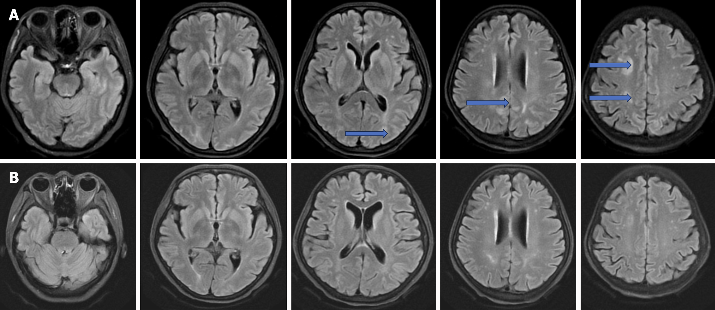
Figure 3 Head magnetic resonance imaging of the liver transplant patient revealed significant recovery in brain white matter damage on postoperative day 22.
A: Scattered areas in the right frontal lobe and bilateral parietal cortex showed slightly decreased T1 signals and slightly increased T2 signals, as indicated by the blue arrows. No significant diffusion-restricted changes were observed on diffusion-weighted imaging (DWI). Specks and flaky abnormal signal shadows were detected beneath the cerebral cortex and near the lateral ventricles on both sides. T1WI displayed low signal intensity, while T2WI and fluid attenuated inversion recovery images (FLAIR) showed high signal intensity. No abnormal high signals were evident on DWI; B: Patchy T1 and T2 signals were noted in the right frontal and bilateral parietal cortex, with no apparent diffusion-restricted changes on DWI or notable abnormal enhancement post-contrast. Specks and flaky abnormal signal shadows were observed beneath the cerebral cortex and near the lateral ventricles on both sides. T1WI showed low signal intensity, whereas both T2WI and FLAIR revealed high signal intensity, with no abnormal high signals detected by DWI.
- Citation: Gong Y. Calcineurin inhibitors-related posterior reversible encephalopathy syndrome in liver transplant recipients: Three case reports and review of literature. World J Hepatol 2024; 16(9): 1297-1307
- URL: https://www.wjgnet.com/1948-5182/full/v16/i9/1297.htm
- DOI: https://dx.doi.org/10.4254/wjh.v16.i9.1297


