Copyright
©The Author(s) 2024.
World J Hepatol. Sep 27, 2024; 16(9): 1297-1307
Published online Sep 27, 2024. doi: 10.4254/wjh.v16.i9.1297
Published online Sep 27, 2024. doi: 10.4254/wjh.v16.i9.1297
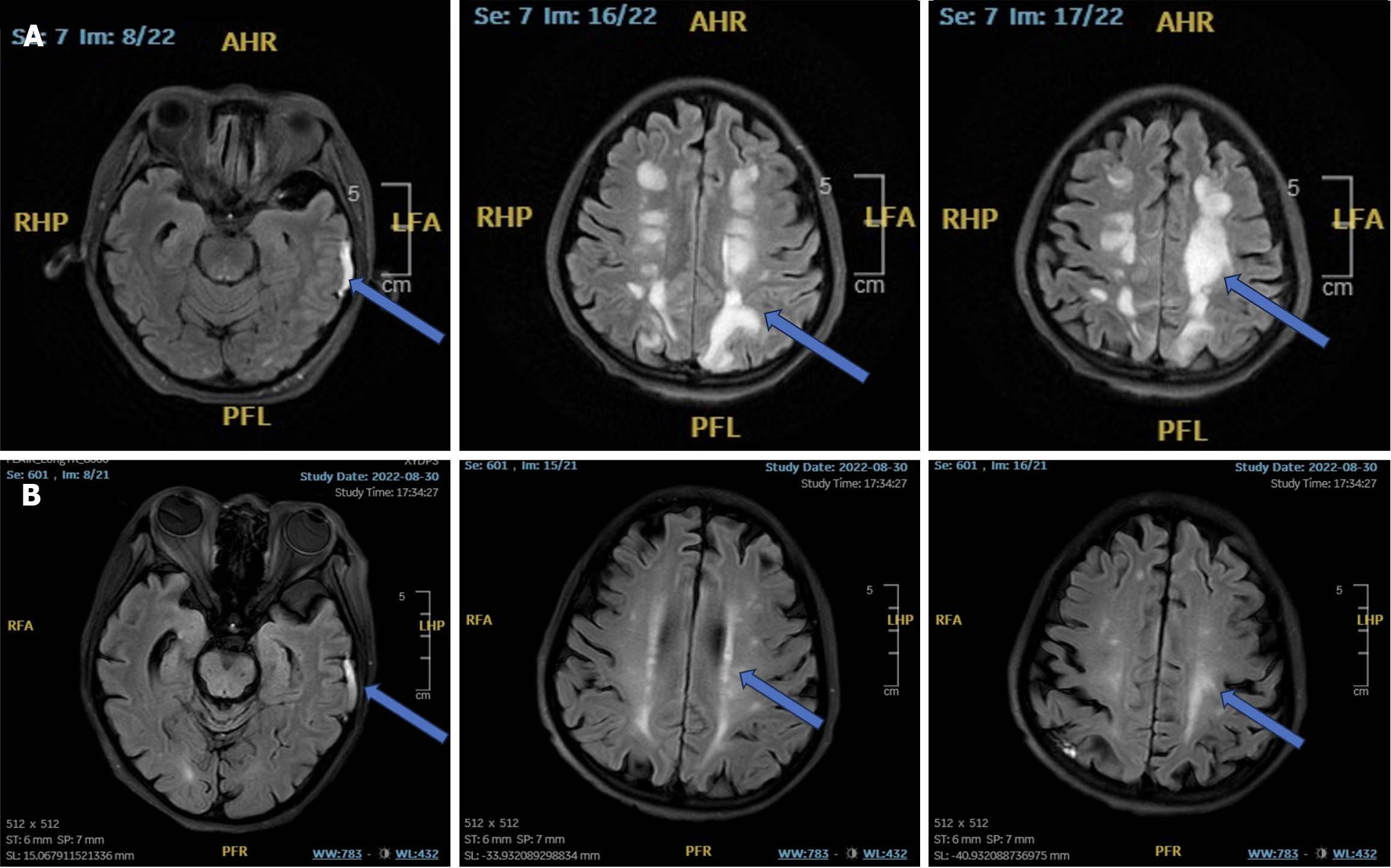
Figure 2 The white matter lesions in the patient’s brain showed gradual resolution over 90 days.
A: Initially, multiple areas with low signal on T1-weighted images (T1WI) and high signal on T2-WI fluid attenuated inversion recovery images (FLAIR) were observed bilaterally beneath the cerebral cortex, in the corona radiata, and the centrum semiovale, as indicated by the blue arrows. No significant abnormalities were noted on diffusion-weighted imaging (DWI). Curvilinear high signal shadows on T1WI and T2WI-FLAIR were also observed in the left temporal subdural region; B: Later, the blue arrows highlight multiple linear regions with low T1WI signal and high T2WI-FLAIR signal, with no evident abnormalities on DWI, along both sides of the cerebral cortex, corona radiata, centrum semiovale, lateral ventricle, and left basal ganglia. Widening of the fluid space was noted under the left medial temporal pole. AHR: Anterior to horizontal right; PFL: Posterior to frontal left; LFA: Left frontal anterior; RHF: Right horizontal frontal.
- Citation: Gong Y. Calcineurin inhibitors-related posterior reversible encephalopathy syndrome in liver transplant recipients: Three case reports and review of literature. World J Hepatol 2024; 16(9): 1297-1307
- URL: https://www.wjgnet.com/1948-5182/full/v16/i9/1297.htm
- DOI: https://dx.doi.org/10.4254/wjh.v16.i9.1297


