Copyright
©The Author(s) 2024.
World J Hepatol. Sep 27, 2024; 16(9): 1297-1307
Published online Sep 27, 2024. doi: 10.4254/wjh.v16.i9.1297
Published online Sep 27, 2024. doi: 10.4254/wjh.v16.i9.1297
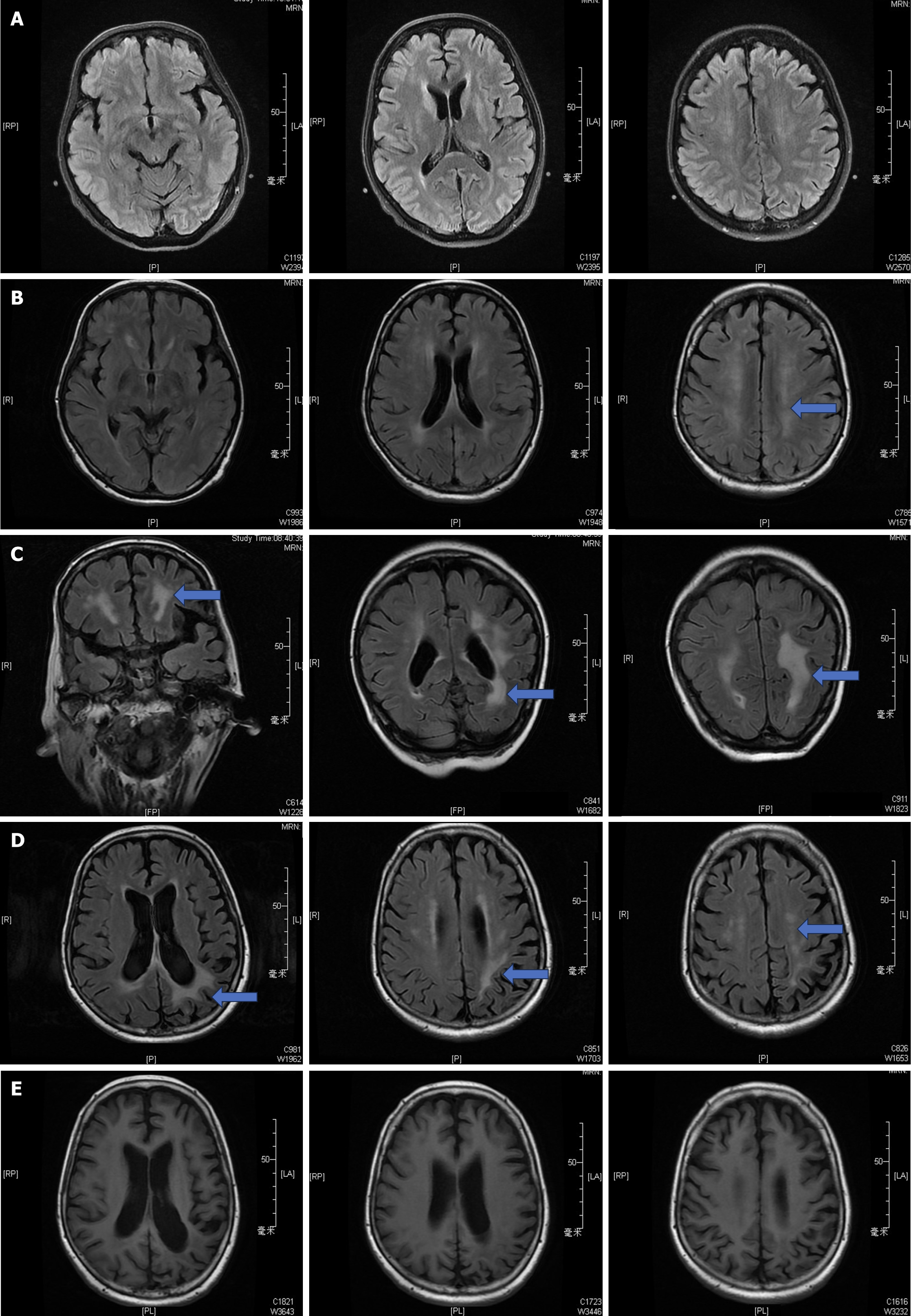
Figure 1 Cranial magnetic resonance imaging findings in patients monitored long-term (2016–2019).
A: On June 15th, 2016 [postoperative day (POD) 9], the cerebral magnetic resonance imaging (MRI) showed ischemic lesions in the bilateral frontal lobes, without noticeable white matter involvement; B: On June 27th, 2016 (POD 22), the MRI revealed a linear high signal in the left posterior parietal region on diffusion-weighted imaging and patchy high signals in the white matter and pons around both sides of the ventricles in fluid attenuated inversion recovery sequences; C: On August 12th, 2016 (POD 66), the MRI continued to show abnormal signals in the white matter surrounding the bilateral ventricles, with low signal intensity on T1-weighted images and slightly elevated signal intensity on T2-weighted images; D: On May 11th, 2018, the MRI still displayed abnormal signals in the white matter around the bilateral ventricles, with low signal intensity on T1-weighted images and slightly elevated intensity on T2-weighted images; E: On May 23th, 2019, the MRI showed no significant abnormalities in the white matter on T1-weighted and T2-weighted images.
- Citation: Gong Y. Calcineurin inhibitors-related posterior reversible encephalopathy syndrome in liver transplant recipients: Three case reports and review of literature. World J Hepatol 2024; 16(9): 1297-1307
- URL: https://www.wjgnet.com/1948-5182/full/v16/i9/1297.htm
- DOI: https://dx.doi.org/10.4254/wjh.v16.i9.1297


