Copyright
©The Author(s) 2024.
World J Hepatol. Sep 27, 2024; 16(9): 1289-1296
Published online Sep 27, 2024. doi: 10.4254/wjh.v16.i9.1289
Published online Sep 27, 2024. doi: 10.4254/wjh.v16.i9.1289
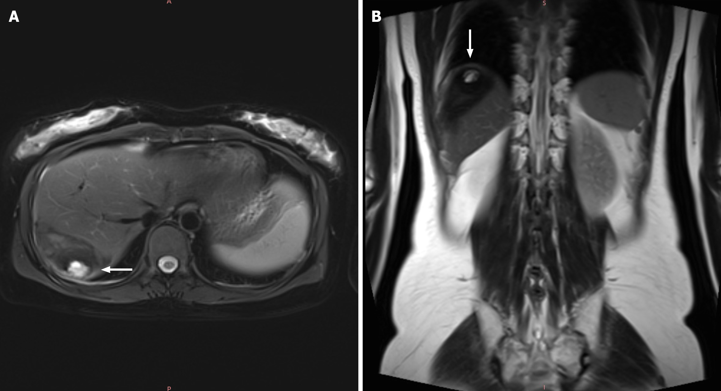
Figure 3 Magnetic resonance imaging (October 2, 2023).
A: Axial scan: The high signal (white arrow) was seemingly under the capsule of liver; B: Coronal scan: The lesion (white arrow) was closer to diaphragm.
- Citation: Yang XC, Fang M, Peng YG, Wang L, Ju R. Hepatic ectopic pregnancy with hemorrhage secondary diaphragmatic adhesion: A case report. World J Hepatol 2024; 16(9): 1289-1296
- URL: https://www.wjgnet.com/1948-5182/full/v16/i9/1289.htm
- DOI: https://dx.doi.org/10.4254/wjh.v16.i9.1289


