Copyright
©The Author(s) 2024.
World J Hepatol. Sep 27, 2024; 16(9): 1211-1228
Published online Sep 27, 2024. doi: 10.4254/wjh.v16.i9.1211
Published online Sep 27, 2024. doi: 10.4254/wjh.v16.i9.1211
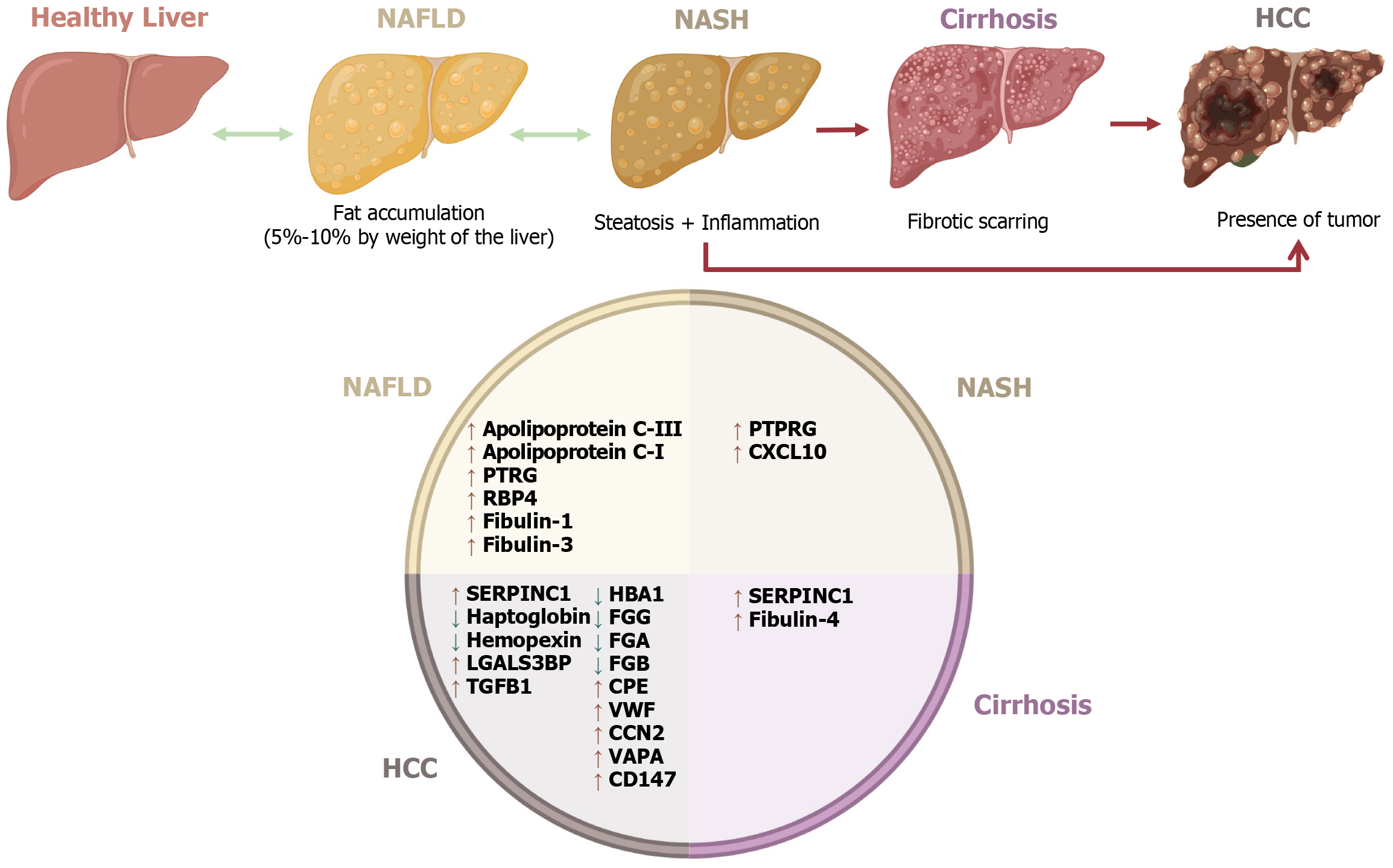
Figure 3 Extracellular vesicle protein expression in different chronic liver disease stages.
The image illustrates the progression of liver disease from a healthy liver to nonalcoholic fatty liver disease (NAFLD), nonalcoholic steatohepatitis (NASH), cirrhosis, and hepatocellular carcinoma (HCC). The circular section lists proteins that increase (↑) or decrease (↓) in each stage: In NAFLD, apolipoprotein C-III (APOC3), apolipoprotein C-I (APOC1), prothymosin alpha (PTMA), retinol-binding protein 4 (RBP4), fibulin-1 (FBLN1), and fibulin-3 (FBLN3); in NASH, protein tyrosine phosphatase receptor type G (PTPRG) and C-X-C motif chemokine ligand 10 (CXCL10); in cirrhosis, serpin family C member 1 (SERPINC1) and fibulin-4 (FBLN4); in HCC, hemoglobin subunit alpha 1 (HBA1), fibrinogen gamma chain (FGC), fibrinogen alpha chain (FGA), fibrinogen beta chain (FGB), von Willebrand factor (VWF), CCN family member 2 (CCN2), vesicle-associated membrane protein associated protein A (VAPA), cluster of differentiation (CD) 147, transforming growth factor beta 1 (TGFB1), galectin-3-binding protein (LGALS3BP), haptoglobin (HP), and hemopexin (HPX). Created in BioRender.com.
- Citation: Montoya-Buelna M, Ramirez-Lopez IG, San Juan-Garcia CA, Garcia-Regalado JJ, Millan-Sanchez MS, de la Cruz-Mosso U, Haramati J, Pereira-Suarez AL, Macias-Barragan J. Contribution of extracellular vesicles to steatosis-related liver disease and their therapeutic potential. World J Hepatol 2024; 16(9): 1211-1228
- URL: https://www.wjgnet.com/1948-5182/full/v16/i9/1211.htm
- DOI: https://dx.doi.org/10.4254/wjh.v16.i9.1211


