Copyright
©The Author(s) 2024.
World J Hepatol. May 27, 2024; 16(5): 751-765
Published online May 27, 2024. doi: 10.4254/wjh.v16.i5.751
Published online May 27, 2024. doi: 10.4254/wjh.v16.i5.751
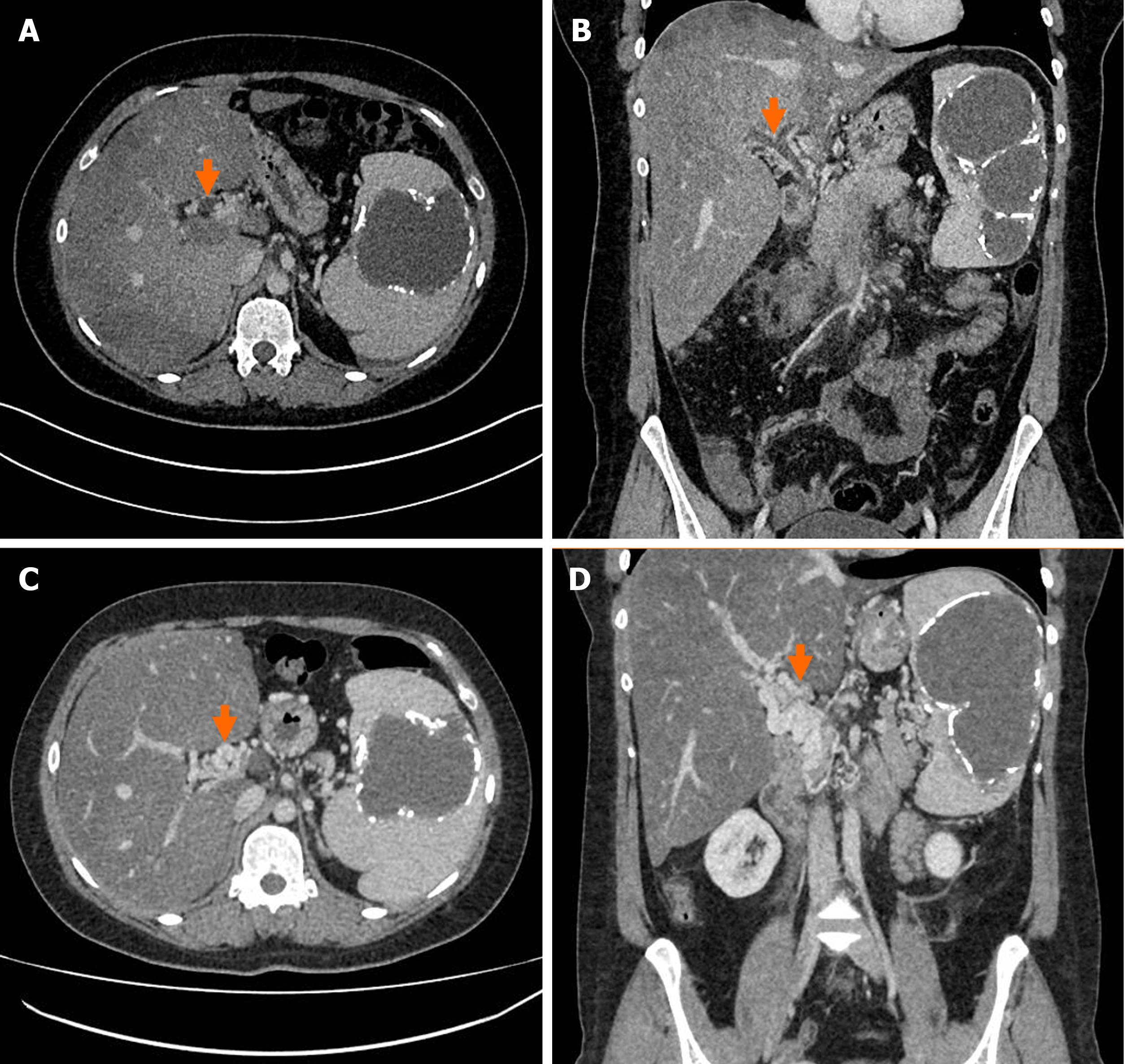
Figure 1 Cross-sectional images of recent portal vein thrombosis and subsequent cavernoma formation.
A: Axial computed tomography demonstrating acute portal vein thrombosis (Orange arrow) with altered hepatic parenchymal attenuation secondary to ischaemia; there is an incidental large splenic cyst; B: Coronal computed tomography demonstrating acute portal vein thrombosis (Orange arrow) with altered hepatic parenchymal attenuation secondary to ischaemia; there is an incidental large splenic cyst; C: Axial computed tomography 6 months later in the same patient, demonstrating formation of portal vein cavernoma (Orange arrow); D: Coronal computed tomography 6 months later in the same patient, demonstrating formation of portal vein cavernoma (Orange arrow).
- Citation: Willington AJ, Tripathi D. Current concepts in the management of non-cirrhotic non-malignant portal vein thrombosis. World J Hepatol 2024; 16(5): 751-765
- URL: https://www.wjgnet.com/1948-5182/full/v16/i5/751.htm
- DOI: https://dx.doi.org/10.4254/wjh.v16.i5.751


