Copyright
©The Author(s) 2024.
World J Hepatol. Mar 27, 2024; 16(3): 316-330
Published online Mar 27, 2024. doi: 10.4254/wjh.v16.i3.316
Published online Mar 27, 2024. doi: 10.4254/wjh.v16.i3.316
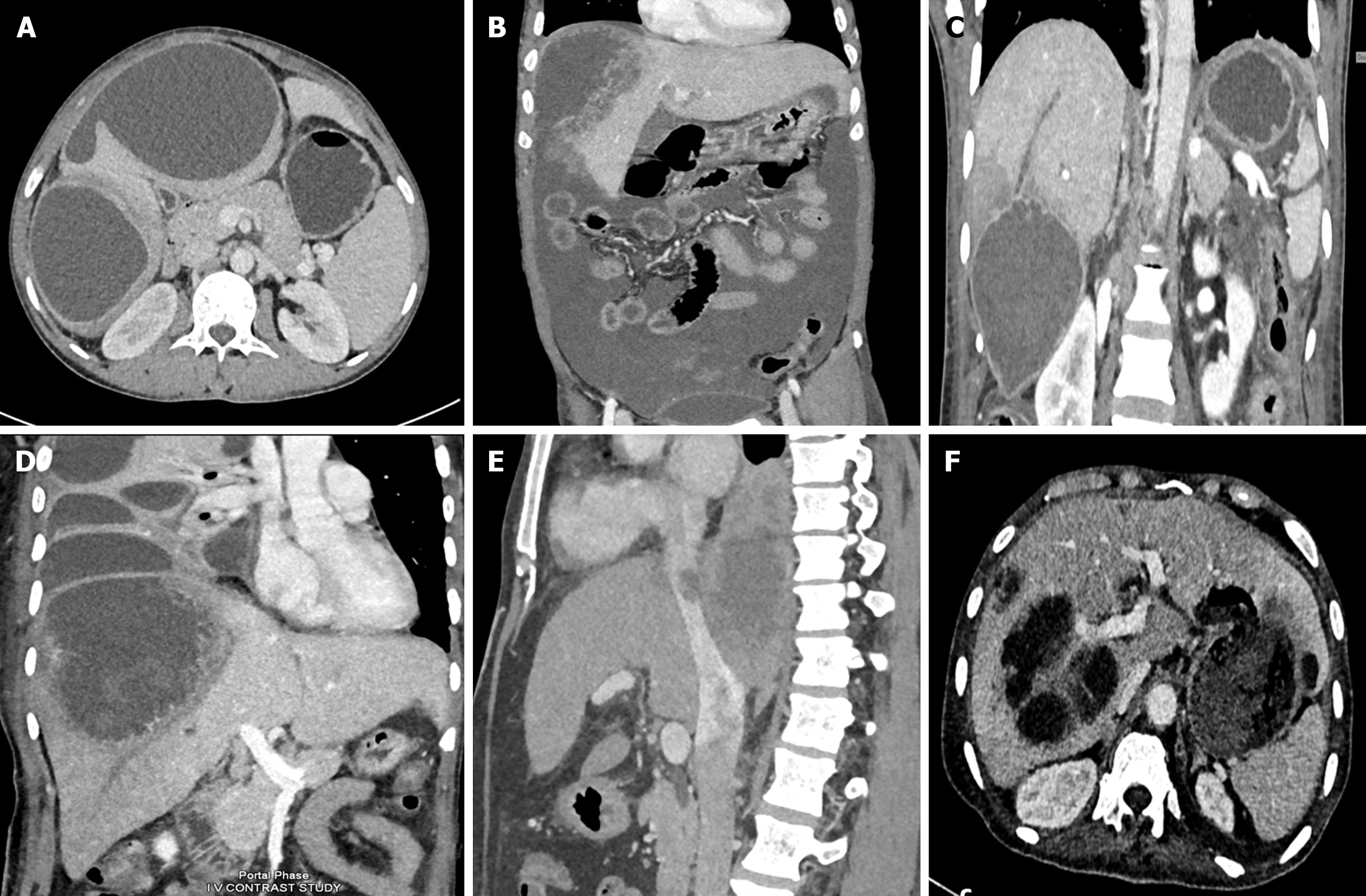
Figure 2 Computed tomographic scans showing various spectrum of complicated amebic liver abscess.
A: Axial computed tomography (CT) scan showing a contained rupture of an abscess with localized fluid collection in the perihepatic area. There is a thin enhancing rim around the abscess, characteristic of a type II amebic liver abscess (ALA); B: Coronal CT scan of a 54-year-old male showing rupture of the abscess with intraperitoneal fluid collection diffusely spreading throughout the entire peritoneal cavity, indicative of free abscess rupture; C: Coronal contrast-enhanced CT image showing an amebic abscess located in segment VI with long-segment thrombosis in the peripheral branch of the right hepatic vein. Note the triangular hypodense area surrounding the abscess, indicating hypoperfused parenchyma; D: Coronal contrast-enhanced CT image showing a large amebic abscess that has ruptured into the thoracic cavity with loculated pleural fluid collections. Note that the abscess demonstrates ragged edges with indistinct enhancement, characteristic of a type I abscess; E: Sagittal contrast-enhanced venous phase CT image of a patient showing thrombus extending from the ALA into the inferior vena cava; F: Axial CT scan showing a communication of left lobe ALA with the gastric lumen forming a hepatogastric fistula.
- Citation: Kumar R, Patel R, Priyadarshi RN, Narayan R, Maji T, Anand U, Soni JR. Amebic liver abscess: An update. World J Hepatol 2024; 16(3): 316-330
- URL: https://www.wjgnet.com/1948-5182/full/v16/i3/316.htm
- DOI: https://dx.doi.org/10.4254/wjh.v16.i3.316


